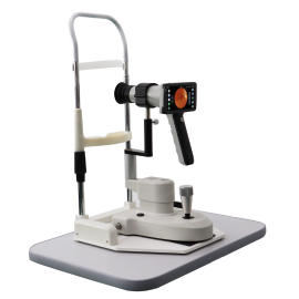

Fundus Cameras
- One-touch operation, automatic shooting.
- No mydriasis, mise au point automatique, and auto exposure
- Automatic chin rest adjustment, automatic pupil detection, automatic working distance adjustment, and automatic alignment
- 24 megapixel camera for high-definition fundus images
- DICOM system connectivity and AI-enabled intelligent systems.
Automatic Fundus Camera
Maxter has launched this fully automated, high-end fundus photography system, utilizing cutting-edge soft exposure technology to capture high-definition fundus images. The highly sensitive touchscreen design allows for flexible and simple operation. Maxter fundus camera can also be integrated with an AI-powered diagnostic system, making your work simpler and more efficient.
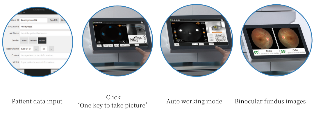
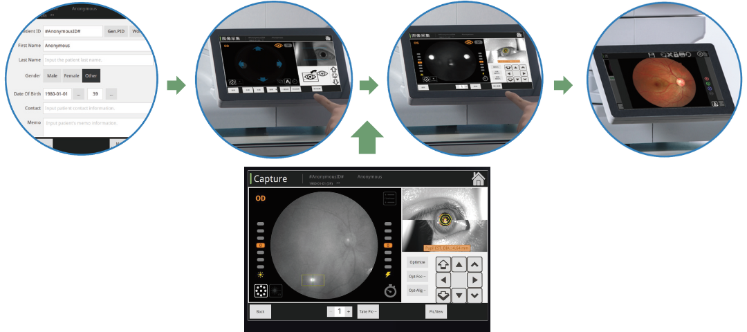
Intelligent Operation -Automatic, Simple and Fast
Automatic Mode
Our fundus camera’s fully automatic working mode can easily achieve fully automatic alignment, focus and automatic photography of both eyes. It does not require any technical skills from the operator and can obtain high-quality photos with one click.
Manual Mode
Our fundus camera can also be switched to manual working mode. When the automatic mode cannot be achieved or when images are collected in a specific fundus area, fundus images can be taken manually by operating the touch screen, which is more convenient than traditional joystick operation.
Soft Exposure-Advanced Optical Technology
Innovative and advanced optical technology allows continuous shooting of the same fundus images of the same patient with almost identical image quality. Soft exposure technology is used in our fundus camera to reduce pupil constriction after exposure.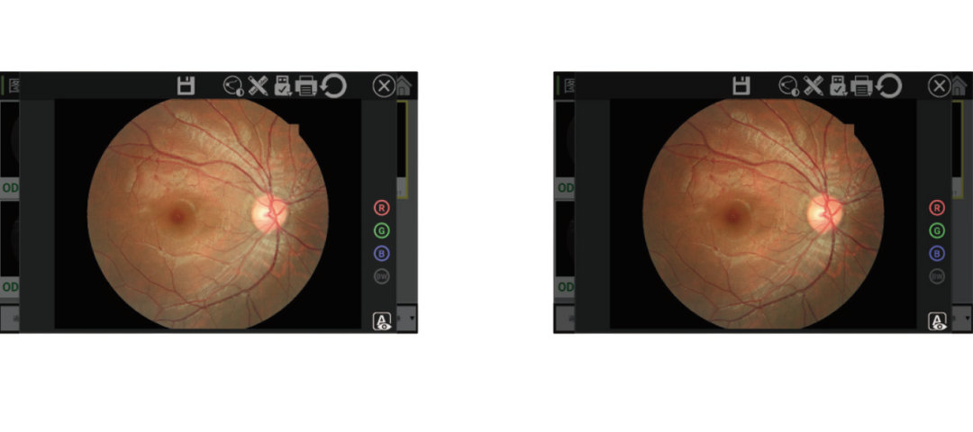
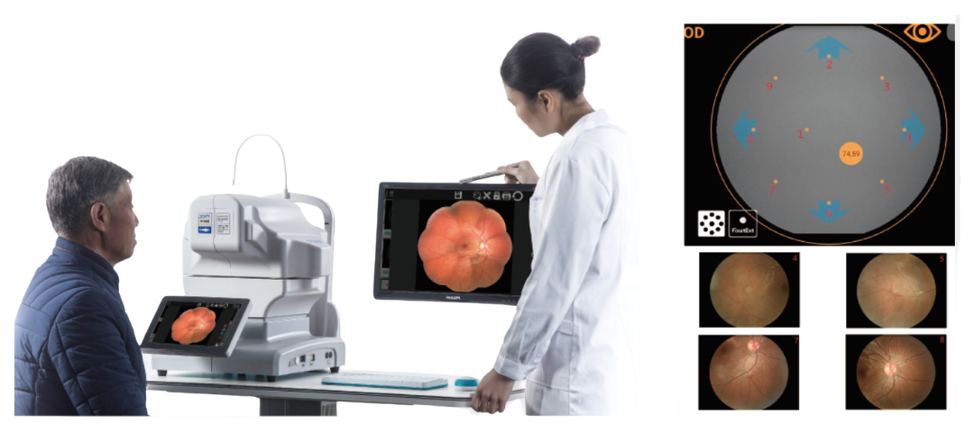
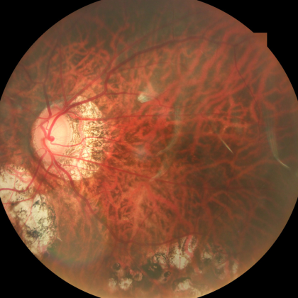
135 Degree Imaging Range
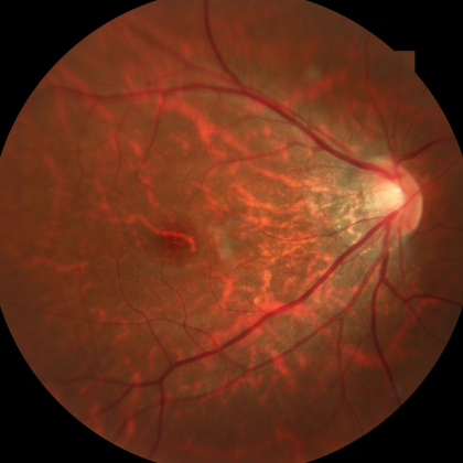
Our automatic fundus camera can produce fundus images of the peripheral retina. In addition to external fixation, internal fixation can be selected arbitrarily. The field of view of a single image can reach 50 degrés. It also features an automatic puzzle function that can reach 135 degrés, providing a wide viewing angle for accurate diagnosis of the fundus situation.
données techniques
| Modes d'acquisition | Non mydriatique/mydriatique
Photographie antérieure Sans rouge(Optionnel) FFA(Facultatif) FAF(Optionnel) |
| Champ de vision | 50° |
| Distance de travail | 35mm |
| Taille minimale des élèves | ≥mm3,3 |
| Mode de mise au point | Automatique/Manuel |
| Mode d'exposition | Automatique/Manuel |
| Opération | Automatique/Manuel |
| Photographie | Appareil photo reflex |
| Résolution | 24Mégapixels |
| Compensation dioptrique | ±25D |
| Fixation | Fixation interne/Fixation externe |
| Assistance à la mise au point automatique | LED infrarouge |
| DICOM 3.0 | Oui |
| Port IA de personnalisation | Oui |
| Dimensions | 38*56*48cm |
| Poids | 27kg |
| Source de courant | 100-240V50/60Hz |
Conception non mydriatique
Minimum Pupil Size of 3.3mm
In small pupil mode, the fundus camera can capture fundus images with a pupil of 3.3mm, which is suitable for a wide range of people and patients who cannot dilate their pupils.

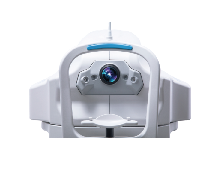
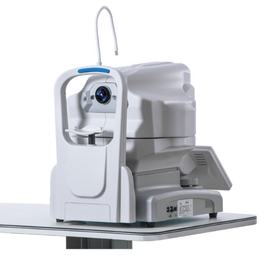
Système à double caméra
High-precision positioning, quickly obtain focus position
Our fundus cameras use a dual-camera system to improve the accuracy and speed of focus, saving operators time, simplifying workflows, and improving efficiency.
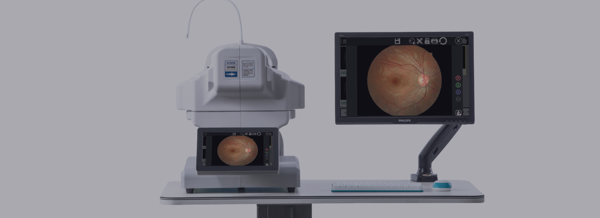
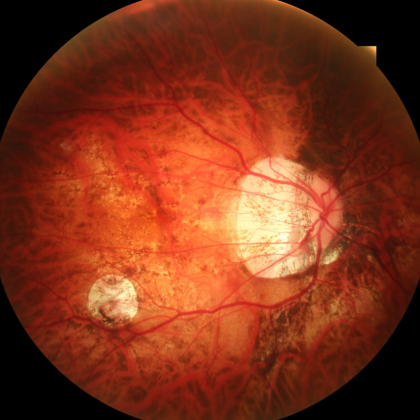
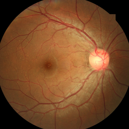
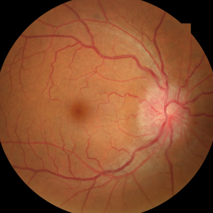
Fundus Cameras at Work
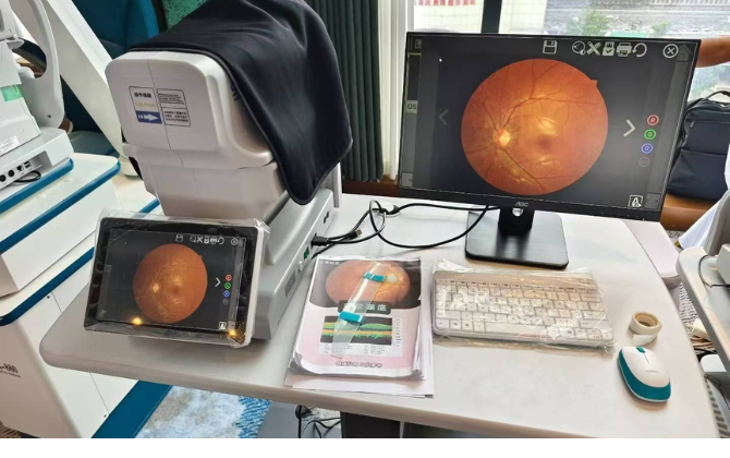
Our non-mydriatic fundus camera is a great partner for doctors, simplifying the daily workflow and improving diagnostic efficiency. Contact us now to get the latest quote!
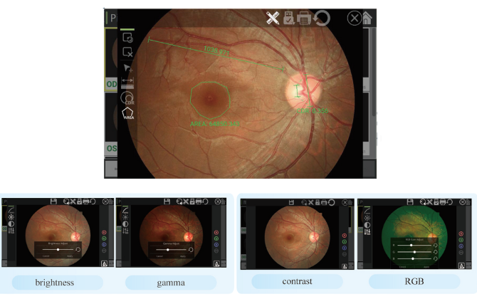
- Powerful image processing capabilities easily determine lesion area and cup-to-disc ratio.
- Image gamma, contrast, luminosité, and color can be easily adjusted to meet your needs.
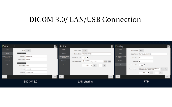
- Supports DICOM protocol, allowing direct connection to hospital PACS and other systems.
- Partage de réseau local: simultaneous recording with the main unit allows real-time display on any computer within the LAN.
- USB expansion for storage.
- FTP remote transfer.

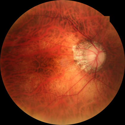
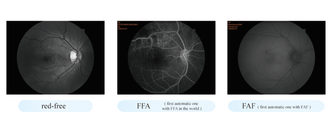
Upgrade Configurations to Present More Details
Our non-mydriatic automatic fundus camera also offers optional Redfree, FFA and FAF functions. After upgrading these functions, it can display more fundus details for more accurate diagnosis.
This fundus camera can be used with or without mydriasis.
Le délai de livraison est dans 3 jours ouvrés après réception de votre paiement. Pour les commandes groupées, don’t hesitate to get in touch with us to determine the exact lead time!
Pour l'instant, Je crains que nous ne puissions pas débiter la carte de crédit! Vous pouvez nous contacter pour plus d'informations sur les modalités de paiement.