- Both table-type and portable-type fundus cameras are available
- Non-mydriatic and safe eye examination
- Rechargeable portable fundus camera
- Mosaik fundus images
- AI diagnosis available
Reliable Fundus Camera Manufacturer
Maxter has been producing fundus cameras for more than 15 years and has an excellent R&D team to ensure the continuous update and iteration of fundus cameras. We are committed to the research and development of fundus cameras without mydriasis. Safe and risk-free fundus examination is our constant pursuit and direction of effort.
Fundus Imaging System
We specialize in fundus imaging and are dedicated to providing simple, fast, and efficient fundus imaging. From portable to fully automated fundus imaging, we are constantly exploring and researching, and updating our models. Please stay tuned to our company for updates on upcoming fundus cameras!


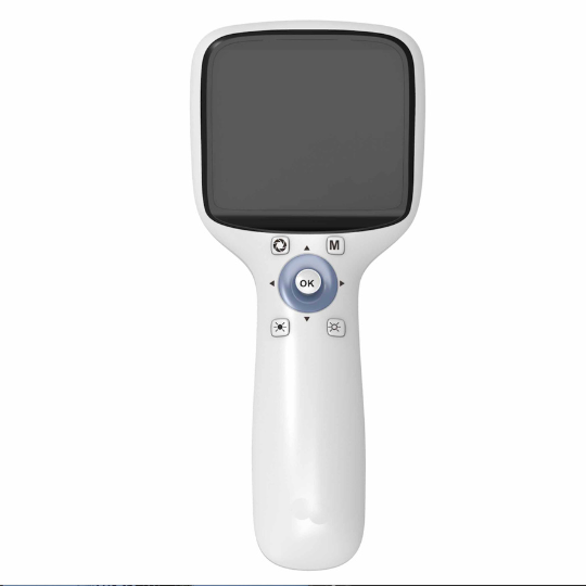
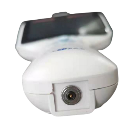
Value Portable Fundus Camera
The PFC01 is our cost-effective portable fundus camera. It can be used for both human and animal eye examinations. It allows for safer fundus examinations without mydriasis, and can capture pupils as small as 2.5mm. The manual focus function provides stability and ease of use. This classic handheld fundus camera, a global bestseller for over a decade, is your affordable option.
-
Our portable fundus camera has a simple design, flexible buttons, supports wireless connection to mobile devices, supports Windows and IOS systems, and easily realizes data transmission.
-
Innovative contact charging design makes charging easier, better protects the charging port, and extends the life of the data cable
-
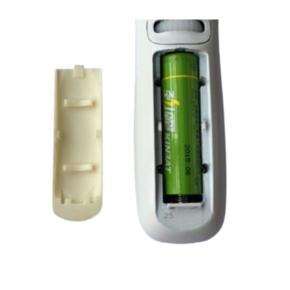
Our fundus camera adopts rechargeable design and lithium battery power supply, which is easy to carry and can be used for fundus examination anytime and anywhere. The battery model is 18650, which is universal and easy to buy.
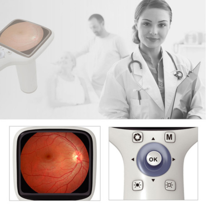
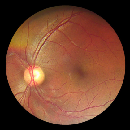
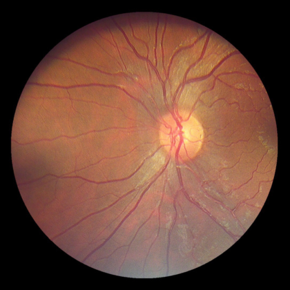
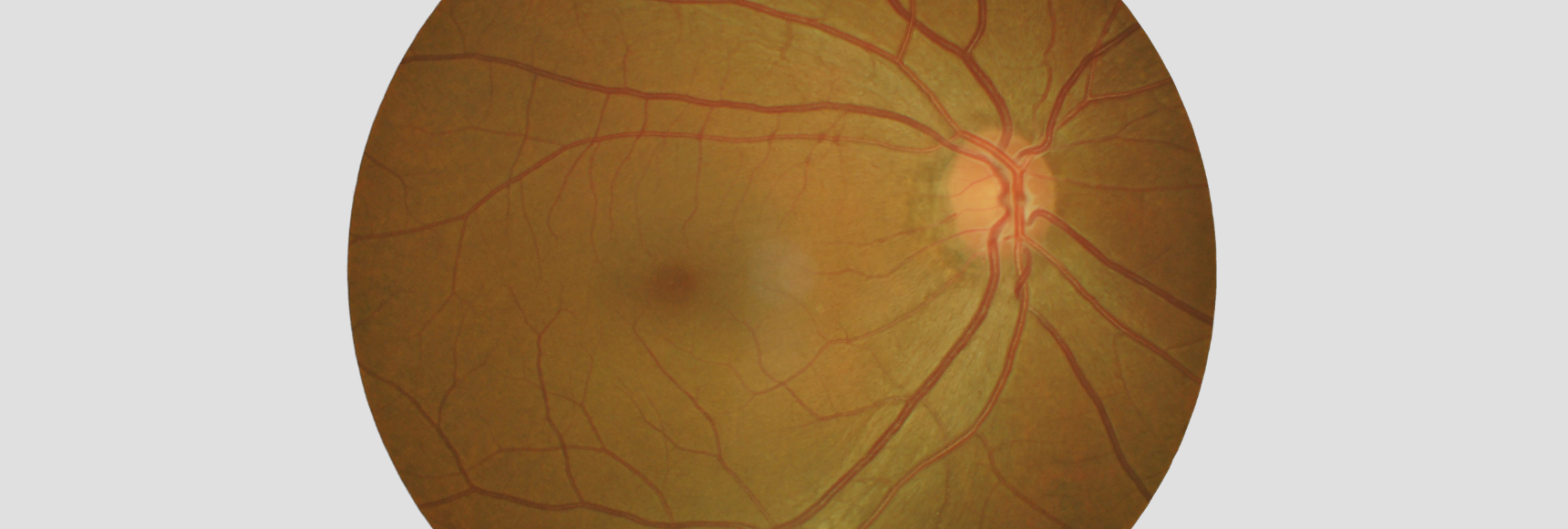
Fundus Images
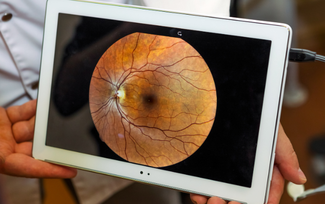
Our fundus camera has a 45-degree field of view, allowing easy acquisition of high-definition fundus images. It also comes with free Apple software that allows for wireless real-time transmission to mobile devices.
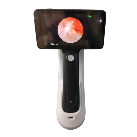
Premium Portable Fundus Camera
The PFC11 is our premium portable fundus camera. It features five internal fixation lights, 18 megapixels, an Android operating system, and a built-in medical record management system. It comes with a lightweight and durable carrying case, allowing you to perform fundus examinations anytime, anywhere. Now, contact us to buy your own mobile fundus camera!
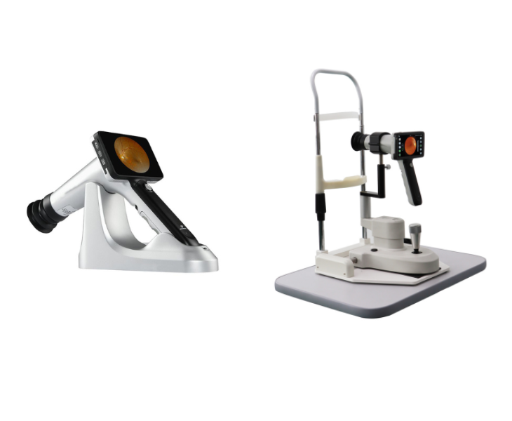
50° Field of View
The premium portable fundus camera provides a 50-degree field of view, allowing you to see the fundus in a wider range. It supports AI intelligent diagnosis, autofocus, and automatic photo taking. The optional bracket instantly transforms it into a desktop fundus camera.
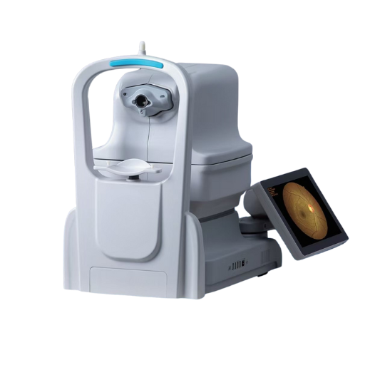
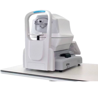
5 Megapixels Fundus Camera
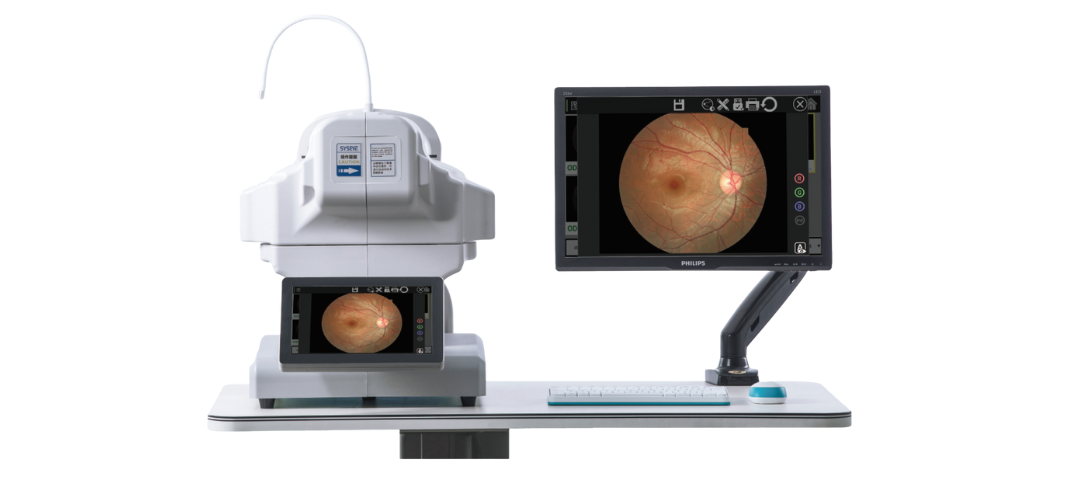
We have launched this 3100m fully automatic non-mydriatic fundus camera, which uses a dual-camera acquisition system and can capture the smallest non-mydriatic pupil of up to 2.8mm. This fundus camera features soft exposure technology, which reduces exposure brightness to increase patient comfort during the examination.
![]()
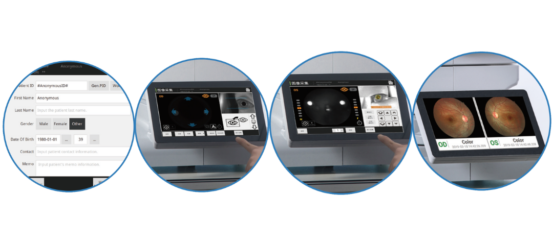
Automatic Working Mode
The 3100m non-mydriatic fundus camera has automatic and manual working modes. In automatic mode, you only need to create a patient profile, and you can automatically adjust the chin rest, automatically focus, automatically align, and automatically take pictures with one click.
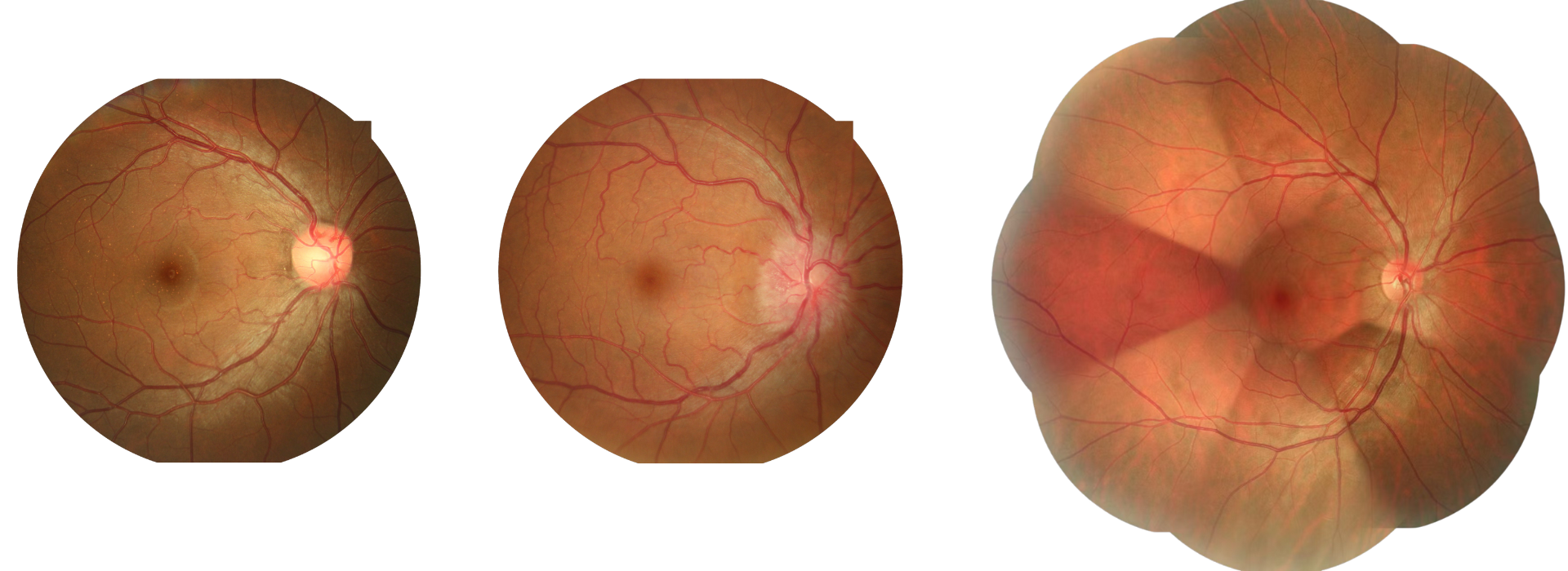
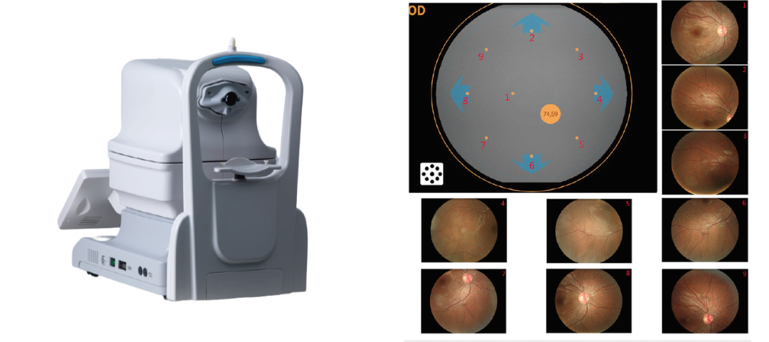
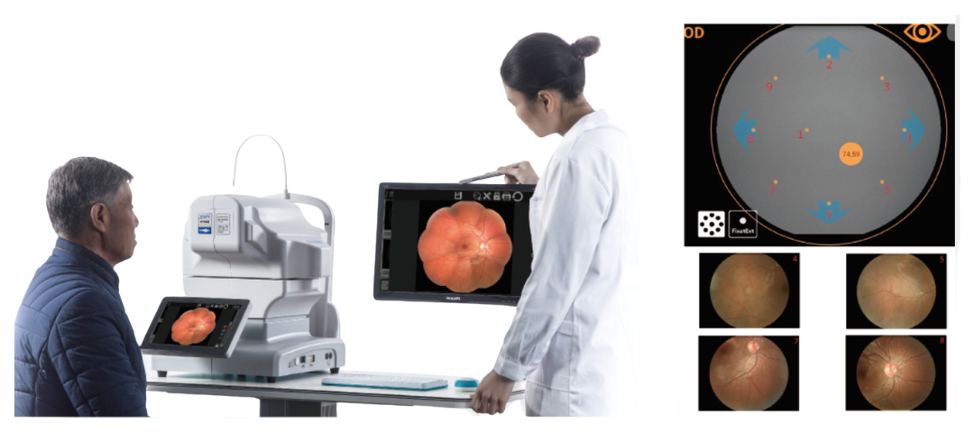
135 Degree Wide Field
9 fundus images at 50 degrees can be combined into a 135-degree ultra-wide-angle to see the entire fundus. The fundus camera has 9 target fixations, and more fixations are distributed between the 9 internal fixations shown.
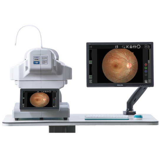
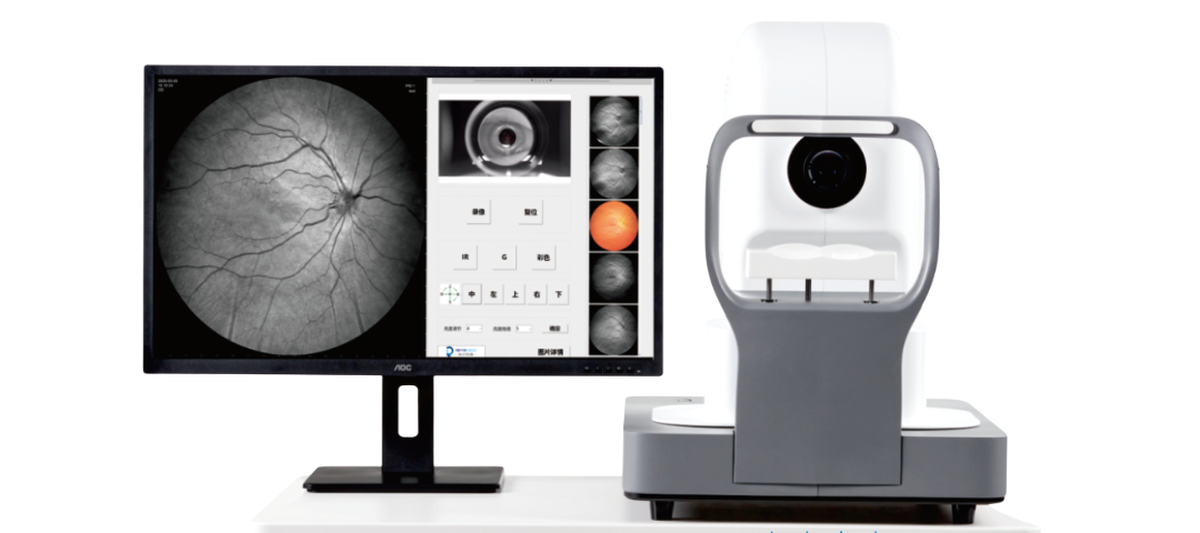
24 Megapixels Fundus Camera
This is our versatile, non-mydriatic, fully automatic fundus camera. Its 24-megapixel camera captures every detail of the fundus in high definition. It has a 50-degree field of view and supports both mydriatic and non-mydriatic modes.
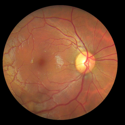
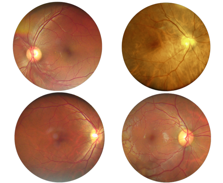
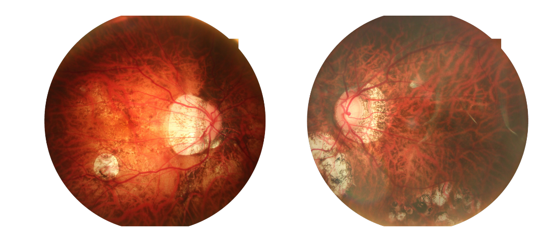
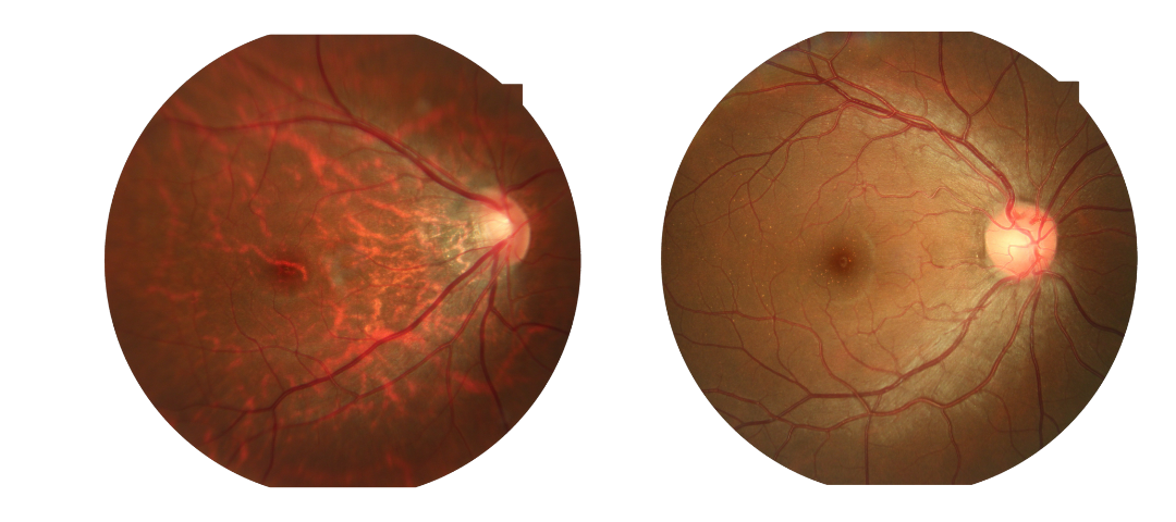
High-definition Fundus Image in Every Detail
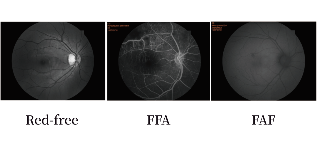
Fundus Camera with FFA
This 3100 fully automatic fundus camera has optional red-free, FFA, FAF, and other functions. It can capture color fundus images and can also meet the needs of fluorescence angiography. It is a cost-effective fundus camera.
One Click to Large Field View up to 135°
With an intelligent medical record management system and one-touch operation mode, our fully automatic fundus camera offers more fundus examination functions, streamlining your workflow and improving efficiency. Join us to explore more wonders of the fundus!
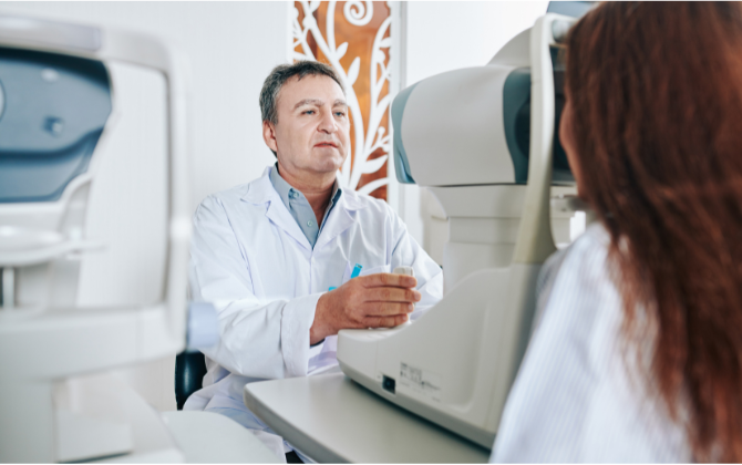
Non-Mydriatic Fundus Camera Applications
Our modern fundus cameras digitize fundus data. This allows for archiving, storage, sharing, and remote consultation. Furthermore, they can be integrated with AI-powered intelligent diagnostic systems to provide reliable diagnostic results.
With the advancement of modern eye diseases and the increasing popularity of laser ophthalmic surgery, postoperative follow-up monitoring is an essential component of postoperative recovery. We can provide customized postoperative follow-up solutions.
Technical Parameter Comparison
Portable Fundus Camera Parameter Comparison
| Portable Fundus Camera | PFC01 | PFC11 |
| FOV | 45 degree | 50 degree |
| Flash mode | White LED/ Near Infrared LED | White LED/ Near Infrared LED |
| Resolution | 1920*1080 | 12 Megapixels |
| Min. pupil | 2.5mm | 2.5mm |
| Focus | Manual | Maunal/Auto |
| Fixation | No | 5 Internal Fixations |
| Screen | 3.5-inch TFT colored screen | 4.7-inch Multi-touch Screen |
| Format | JPEG | Color image, Monochrome image, Infrared image |
| Inerface | Micro USB | USB3.0 |
| Image transfer | Micro USB, WIFI | USB3.0,WIFI, Bluetooth,SD, Type C |
| Storage | Micro SD card, maximum to 32G | 4G+64G |
| Power | Li-on rechargeable battery 3.6V/3400mAh | Li-on rechargeable battery 3.7V/3500mAh |
| Net weight | 400g | 600g |
| Size | 19*10*21cm | 41*25*35mm |
Auto Fundus Camera Parameter Comparison
| Auto Fundus Camera | 3100 | 3100m |
| Acquisition Modes | Non-Mydriatic/Mydriatic
Anterior Photography Red-free(Optioanl) FFA(Optional) FAF(Optioanl) |
Non-Mydriatic/Mydriatic |
| Field of View | 50° | 50° |
| Working Distance | 35mm | 13mm |
| Minimum Pupil Size | ≥mm3.3 | ≥2.8mm |
| Focus Mode | Auto/Manual | Auto/Manual |
| Exposure Mode | Auto/Manual | Auto/Manual |
| Operation | Auto/Manual | Auto/Manual |
| Photography | SLR Camera | SLR Camera |
| Resolution | 24Mega Pixels | 5 Mega Pixels |
| Diopter Compensation | ±25D | ±25D |
| Fixation | Internal Fixation/External Fixation | Internal Fixation |
| Autofocus Assistance | Infrared LED | Dual Camera |
| DICOM 3.0 | Yes | Yes |
| Customization AI Port | Yes | Yes |
| Dimensions | 38*56*48cm | 28*56*46cm |
| Weight | 27kg | 21kg |
| Power Supply | 100-240V 50/60Hz | 100-240V 50/60Hz |
-
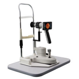
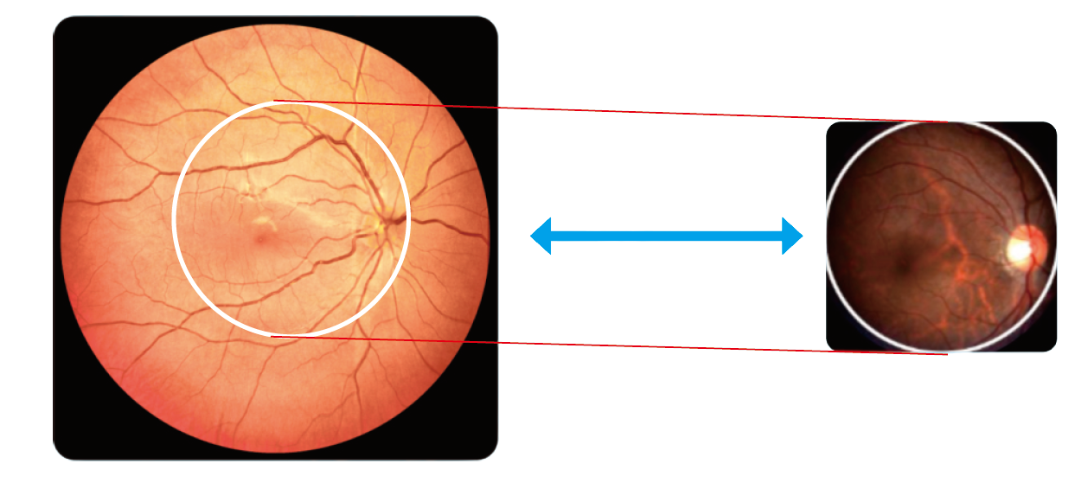
Confocal Laser Scanning
The new generation ultra-widefield laser confocal fundus camera supports non-mydriatic and ultra-widefield imaging. Utilizing a laser confocal system enables fast and precise examinations, enhancing patient comfort, particularly for patients with glaucoma or diabetes who are unsuitable for mydriasis.
Wide-field Retinal Imaging
Our confocal laser fundus cameras support multiple light source options, allowing for easy achievement of ultra-wide-angle field of view of 130° and 168°, and enabling one-click image acquisition. Currently, single-light source viewing is available for choroidal observation, while dual-light source viewing is available for both the retina and choroid. A quad-light source viewing system for both the retina and choroid is coming soon.
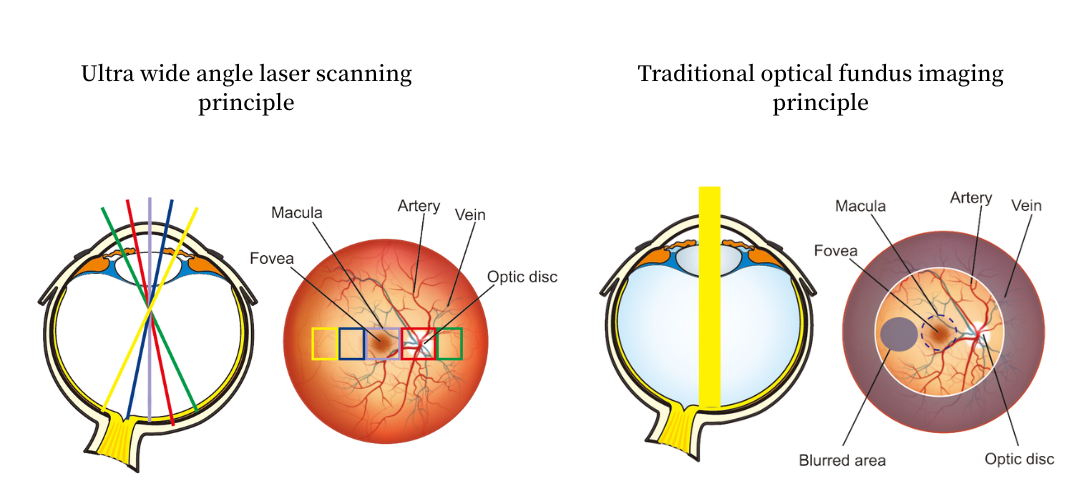
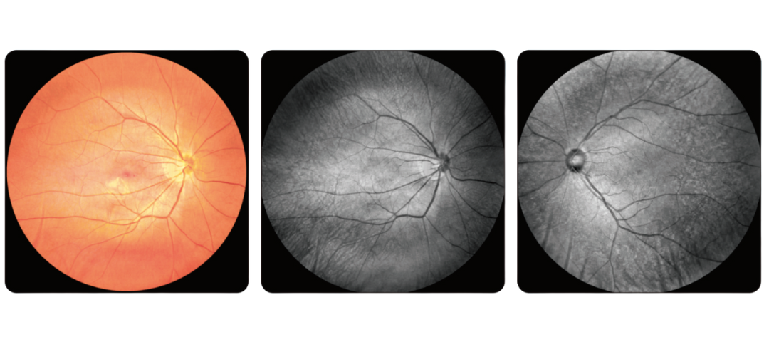
SLO Working Principle
Retinal full-layer photography + Video
The full-layer retinal imaging/videography technology allows more accurate identification of pathological areas, providing a basis for personalized treatment.
Normally, we have products in stock, and we can ship out within 2 working days after receiving your payment! Please contact us for the exact lead time for bulk orders!
Yes, we can offer training if you need! Please get in touch with us for our training video!
Generally, we offer a 1-year warranty for our products. But for some models, we offer a longer warranty period. Contact us for more information about the warranty time!