

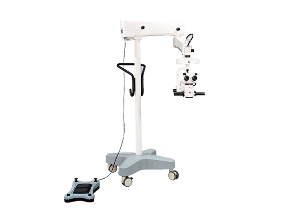


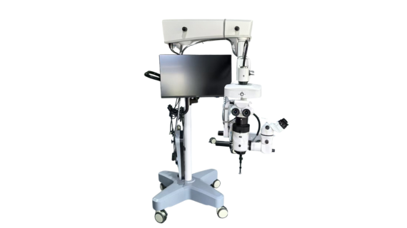
Microscopio operativo ROMS100
Características
- ROMS 100 Utiliza fuente de luz LED coaxial
- El sistema de iluminación está equipado con un dispositivo de protección de la retina, -2° Intensificador Independiente del Reflejo Rojo, Filtro de Infrarrojos
- Sistema X/Y controlable automáticamente, Y el rango móvil de X/Y:50milímetros
- El instrumento adopta un contrapeso de resorte de gas en cada punto fijo
- El rendimiento es fácil y flexible a la vez que estable y confiable
Acerca de nuestro Microscopio oftálmico
Nuestro microscopio oftálmico ROMS100 es aplicable para cirugías del segmento anterior y posterior.. La ruta óptica principal está diseñada con cuatro rutas ópticas y adopta un complejo sistema óptico de aberración cromática.. Con iluminación coaxial y dos fuentes de iluminación LED., Nuestro microscopio quirúrgico proporciona un brillo excelente para procedimientos quirúrgicos..
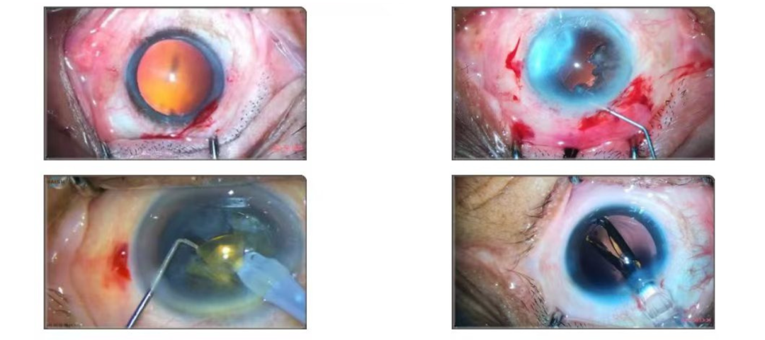
Cirugías del segmento anterior
- Puedes ver claramente las diferentes capas., zonas nubladas, y las relaciones entre los tejidos circundantes del cristalino.
- Limpiar el área quirúrgica: incluyendo incisión quirúrgica, cámara anterior, cámara posterior, etc., para que pueda realizar con precisión cada paso quirúrgico y evitar dañar importantes tejidos circundantes.
- No hay áreas de sombra obvias dentro del campo de visión quirúrgico para garantizar que pueda observar el sitio quirúrgico de manera integral..
- El color de la imagen debe reflejar la situación real del tejido de la forma más realista posible., para que pueda juzgar con precisión el estado de salud y el grado de lesiones del tejido.
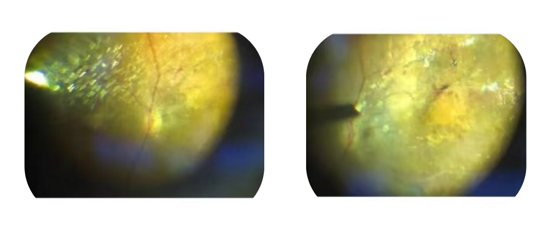
Cirugías del segmento posterior
- Los médicos asistentes pueden realizar cirugías posteriores con alta precisión en condiciones de iluminación de bajo brillo..
- Nuestro microscopio oftálmico proporciona una excelente profundidad de campo con un aumento de 6X a 8X, evitando ajustes frecuentes de enfoque.
- El microscopio oftálmico ROMS100 es bueno para mantener una visualización clara de la retina , especialmente cuando se utilizan sistemas de observación de gran angular de 130 grados.
- Durante el proceso de pelado, El microscopio oftálmico ROMS100 también puede obtener una visualización clara de la capa de película..
- El diseño de 4 Los caminos ópticos garantizan una iluminación suficiente para la visualización y al mismo tiempo evitan el riesgo de exposición fototóxica..
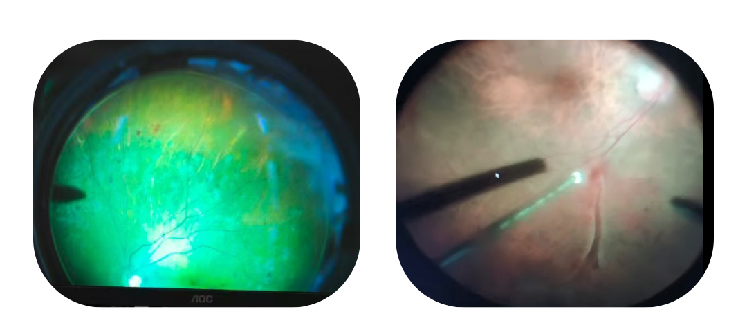
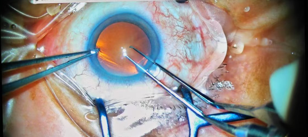
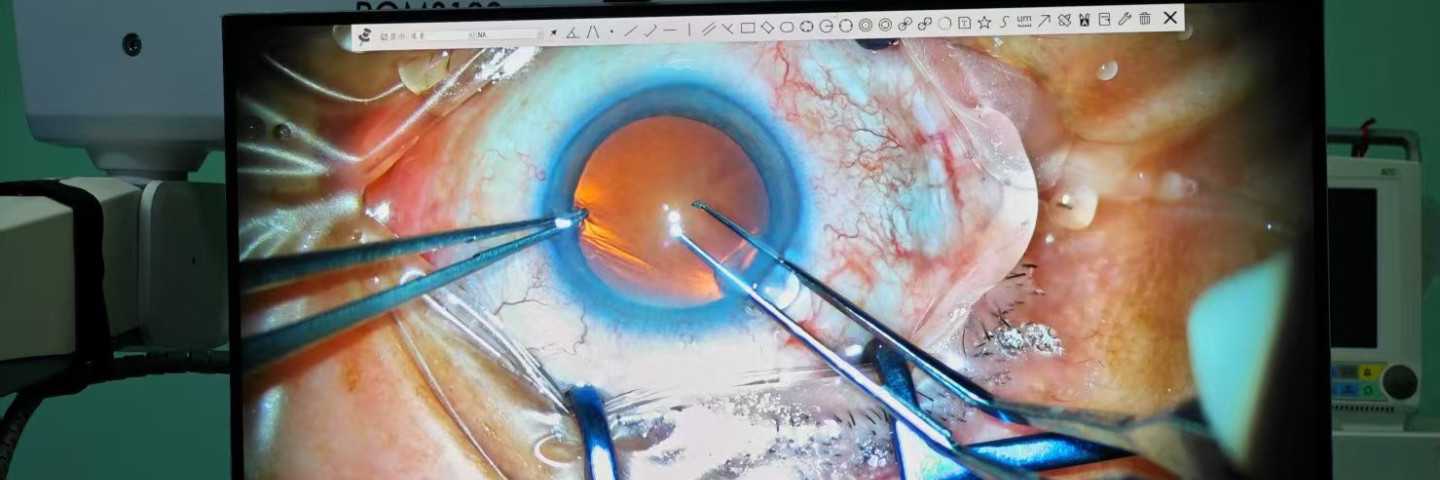
Reflejo rojo superior para cirugías de retina
El reflejo rojo es un componente crucial de los microscopios quirúrgicos oftálmicos.. Su calidad impacta directamente en la capacidad del microscopio para realizar cirugía del segmento posterior.. La pupila está iluminada por la luz roja del microscopio., y se observa la luz roja reflejada. Normalmente, debería aparecer rojo uniforme. El color o la forma anormal pueden indicar cataratas., errores refractivos, o retinopatía. Nuestro microscopio quirúrgico ROMS100 proporciona un reflejo rojo claro y brillante., creando un entorno operativo excelente para la cirugía del segmento posterior.
Iluminación y datos eléctricos
| Luz principal | LED integrado, 3500k, IRC≥95 |
| Iluminación refleja roja | -2°Coaxiales, CONDUJO, 3500K IRC≥90 |
| Entrada de voltaje | 100~240V, 50/60Hz |
| Fusibles | 250V, 10A |
| Normas de seguridad eléctrica | IEC60601-1: 2012, Clase I |
Sistema óptico
| Oculares | 12.5incógnita |
| Rango de ajuste de dioptrías | -7D~+7D |
| Objetivo | F=200mm |
| Distancia de trabajo | 190milímetros |
| Ampliación del sistema óptico principal | ZOOM 6:1 Motorizado |
| Ampliaciones | Óptica Principal : 4.1~24,6X;
Asistente de Óptica:5.4incógnita, 8incógnita, 12.2incógnita |
| Diámetro del campo | Óptica Principal: 8mm ~ 50 mm
Asistente de Óptica: 34.6milímetros, 23milímetros, 14.63milímetros |
| PD para binocular principal | 51milímetros ~ 75 mm |
| PD para binoculares auxiliares | 55milímetros ~ 75 mm |
| Rango de inclinación del binocular principal | 0~180° |
| Enfoque | 50milímetros, Motorizado con reinicio automático |
| Velocidad de enfoque | ≤2 mm/s |
| Filtros | Iluminador principal: Filtro de protección de retina, libre de rojo, GG435 Bloqueo azul;
Iluminador reflejo rojo:20% Atenuación de luz |
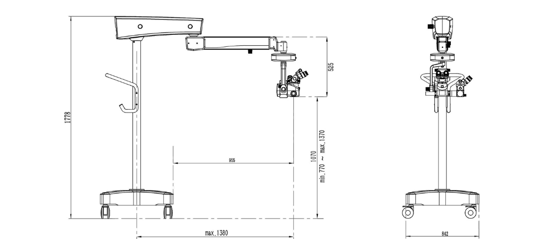
Datos del soporte de suelo
| Ruedas de pie | 4*100milímetros |
| Peso | Aproximadamente 185 kg, Sistema de microscopio completo |
| Rango | Extensión 1380mm(Máx.) |
| Equilibrio | Resorte de gas |
| Unidad XY | 50milímetro*50m m, Motorizado con reinicio automático |
| Velocidad de movimiento XY | ≤2 mm/s |
| Controlador de inclinación | Manual, +15°/-50° |
| Rango de giro | Brazo Horizontal:360°
Brazo de equilibrio:±135° Unidad XY:±135° |
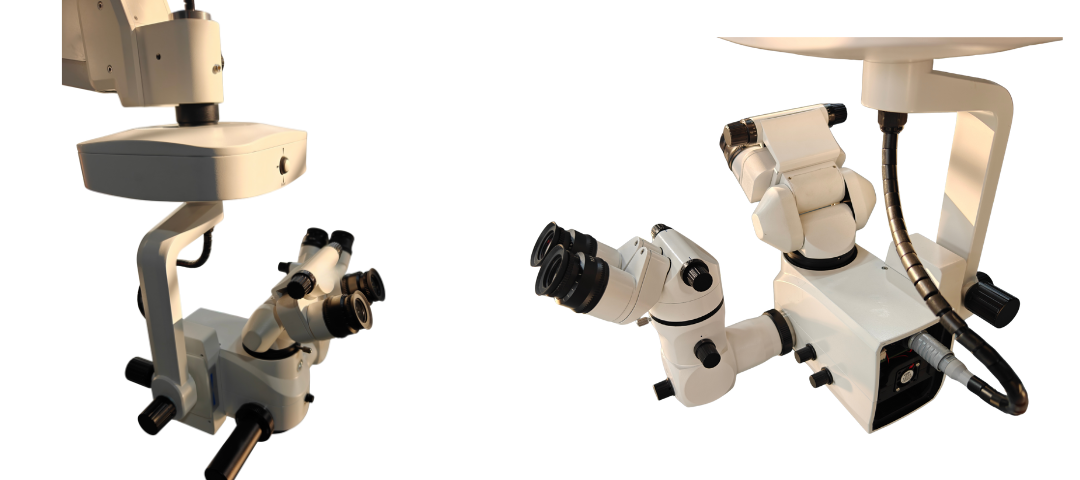
Alcances extensibles para más operadores

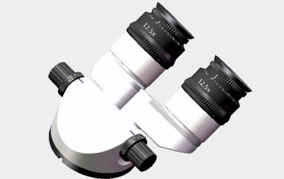
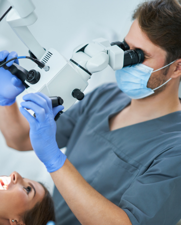
Tubo binocular para 0~180°
El tubo binocular de nuestro microscopio quirúrgico se puede ajustar desde 0 a 180 grados, lo que mejora la comodidad y la flexibilidad a través de una gama aún más amplia de ángulos de visión.
Extensible para 2 Operadores
Hay disponible un microscopio binocular auxiliar con una inclinación de 45 grados., que funciona en una cómoda posición estándar con un ángulo de visión fijo de 45°.
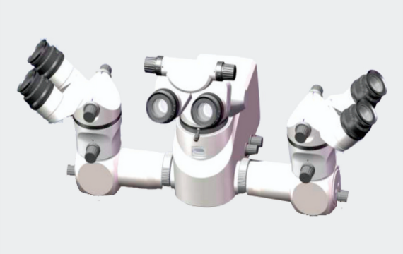
Extensible para 3 Operadores
Se pueden montar dos telescopios auxiliares a ambos lados del microscopio para compartir todo el proceso quirúrgico..
Filtros láser
Los filtros de protección láser opcionales para nuestro microscopio oftálmico brindan una mejor protección para el operador, con un nivel de protección superior a 4. Hay tres filtros láser de longitud de onda disponibles.: 532Nuevo Méjico, 577Nuevo Méjico, y 810nm. Bienvenido a ponerse en contacto con nosotros para obtener más información.!
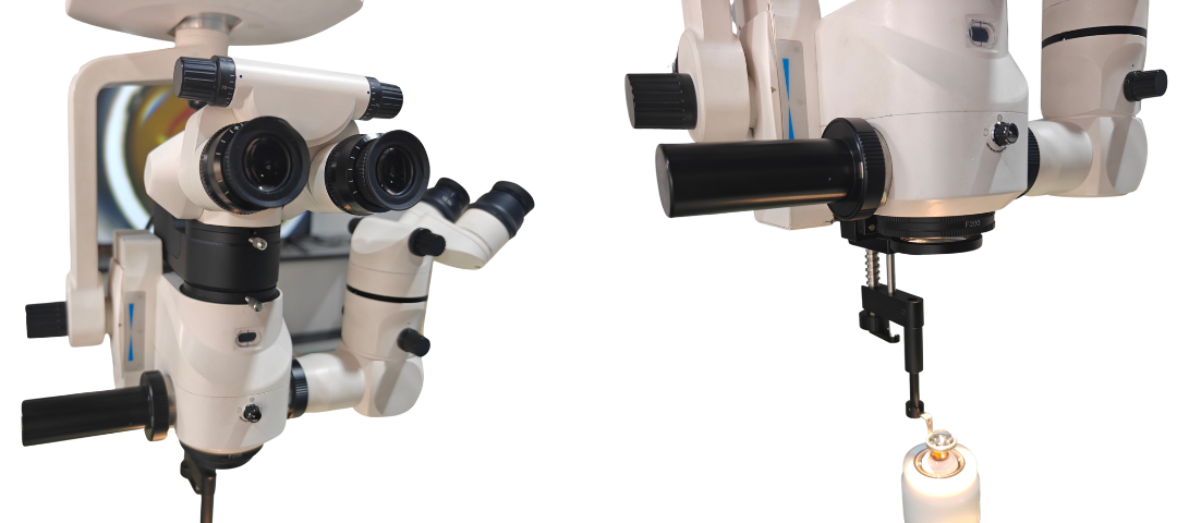
Estructura del microscopio operativo
En la estructura de diseño del microscopio quirúrgico., Hemos considerado plenamente la conveniencia del operador y adoptamos un pedal con cable para garantizar una conexión más estable y un proceso quirúrgico más fluido..
Microscopio compacto y equilibrado
El diseño general presenta el microscopio principal y un microscopio auxiliar., caracterizado por una estructura compacta y equilibrada que facilita un movimiento estable y fácil.
Sistema quirúrgico de retina confiable
Después de una extensa validación clínica, Nuestro microscopio quirúrgico del segmento posterior ha obtenido un amplio reconocimiento y grandes elogios.. Superando las limitaciones tecnológicas y logrando una mayor profundidad de campo, Proporciona una solución superior para la cirugía de retina..
Accesorios (3)
-

El sistema 4K HD integrado es opcional, como sistema de actualización digital para nuestro microscopio quirúrgico ROMS100, que puede equiparse con un monitor de alta definición para visualizar el proceso quirúrgico.
-
Como accesorio del microscopio quirúrgico., El espejo inversor se utiliza junto con el sistema BIOM en cirugía oftálmica clínica para invertir la imagen retiniana invertida formada por el BIOM y obtener una imagen vertical..
-

Nuestro sistema BIOM es un sistema óptico que ayuda en el tratamiento y cirugía oftálmica a través de principios ópticos como la magnificación o la inversión..
Sistema visual 4K actualizable
Nuestro microscopio quirúrgico se puede actualizar al sistema de alta definición 4K, y cuando se conecta a un monitor de alta definición, Todo el proceso quirúrgico se puede observar en tiempo real.. Contáctenos para actualizar su microscopio!
Operante Aplicación de microscopio


El microscopio ROMS100 es el más claro entre las marcas nacionales para cirugía del segmento posterior. Al publicar nuestros artículos sobre investigación del segmento posterior, debemos incluir el microscopio ROMS100 de Maxter Medical en el artículo.–Profe. Yang del Hospital Afiliado de la Universidad Tecnológica de Qilu
El microscopio Maxter tiene la óptica más clara entre las marcas nacionales que he visto.. Es mucho mejor que otras marcas nacionales de microscopios oftálmicos que vi en la 2024 Reunión Anual Nacional de Oftalmología. El reflejo de la luz roja es muy bueno y la profundidad de campo es suficiente para la cirugía del segmento posterior., lo que superó mis expectativas para los microscopios oftálmicos domésticos.– Profe. Él del Hospital del Gobierno Provincial de Hubei.
El microscopio ROMS100 está equipado con un sistema de grabación de vídeo de alta definición 4K, que registra muy bien todo el proceso de nuestra cirugía de cataratas.. Las imágenes son verdaderamente de alta definición., lo cual es conveniente para nosotros utilizar para la enseñanza y la capacitación.–Profe. Dong del hospital Anhui SuChu
El microscopio ROMS100 tiene la mejor resolución que nunca antes había visto en ningún otro microscopio oftálmico de marca china., Es realmente perfecto mientras lo reviso sin que se enciendan las luces.–Profe. Vadim de TMS Rusia