Premium Features
- Optimum optical design provides more detail
- 14mm large spot size design provides a wider field of view
- Ultra-large diopter compensation -7D to +7D meets more needs
- Integrated beam splitter for easier installation
- Integrated digital imaging system simplifies operation
- External yellow filter available
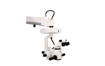
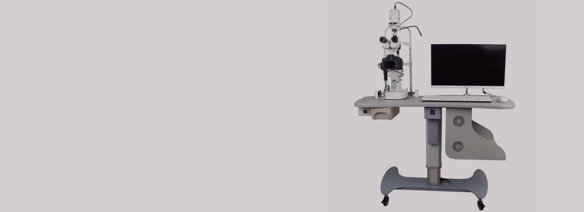
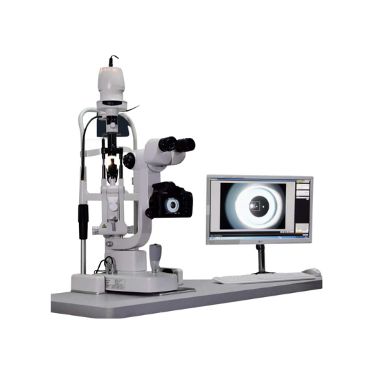
Digital Slit Lamp SL-1911D
This is a premium digital slit-lamp microscope. The large spot size provides a wider field of view and a larger observation area. The broader refractive power compensation range is from -7D to +7D, making it suitable for a wider range of ophthalmologists.
Management System
The SL-1911D digital slit lamp is equipped with professional slit lamp image processing software that records and saves the patient’s eye movements in real-time, establishing a comprehensive tracking record to help you systematically manage medical records.

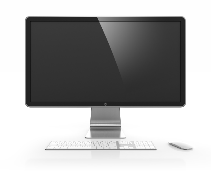
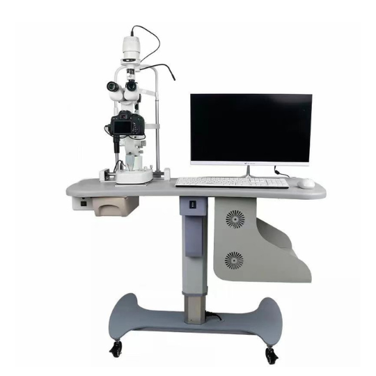
All-in-one Computer
The desktop all-in-one computer, as a digital upgrade accessory for this slit lamp, features an i3-5005u processor, 8GB of RAM, and a 256GB hard drive. Combined with the image processing system, it enables the slit lamp to operate intelligently. Of course, you can also choose the computer configuration according to your needs, and we can upgrade it for you.
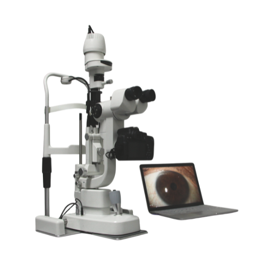
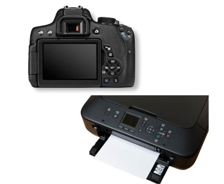
Canon Digital Camera and Printer
The digital slit lamp also comes with a Canon digital camera and a Canon colour inkjet printer for easy high-definition photography and medical record printing.
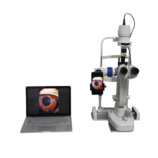
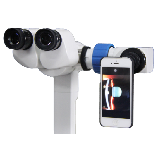
Digital Upgrade and Wide Field of View
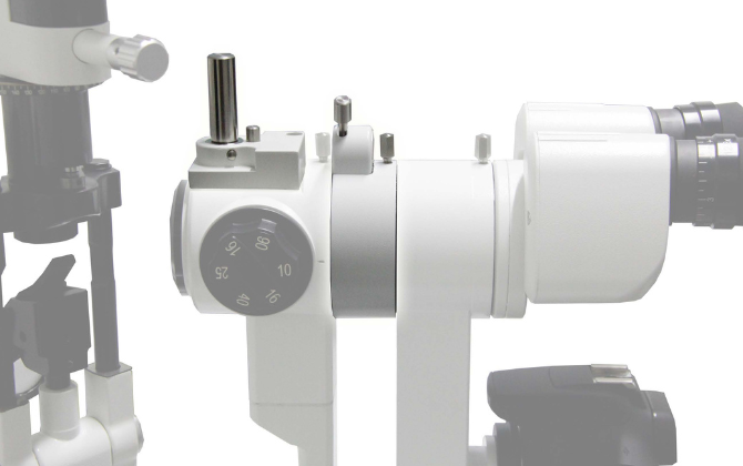
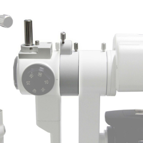
The beam splitter divides the slit lamp light into two beams: one for observation under a microscope and the other for a digital camera. The integrated splitter is more streamlined, easier to install and remove, and simplifies digital upgrades.

The 1911D digital slit lamp features a large 14mm light plate and four types of filters, enabling it to examine various diseases of the anterior segment and providing important information for ophthalmic diagnosis.
Optimum optical design delivers a realistic and three-dimensional observation experience, making it easy to observe and photograph corneal endothelial cells.
The yellow filter is optional for the SL-1911D digital slit lamp, which effectively enhances the contrast between lesions and normal tissues, and improves the accuracy of doctors’ diagnosis. If you require the yellow filter function, you can also contact us to check whether your slit lamp is compatible with it.
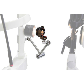
The optional fundus lens holder, when used with a 90D aspherical lens, facilitates easy observation of the fundus.
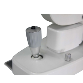
We installed a camera shutter on the joystick. When doctors use the joystick to adjust the focus and find the clearest position, they can quickly press the shutter to take a clear image without having to look for the camera shutter, which saves time and captures the moment.
- Specifications
Optical system
| Microscope type | Galilean Steroscope Microscope |
| Magnification type | Drum Type 5 Magnifications |
| Eyepiece | 12.5X |
| Magnifications | 6X, 10X, 16X, 25X, 40X |
| Field of view | ϕ33mm, ϕ22.5mm, ϕ14mm, ϕ8.8mm, ϕ5.5mm |
| Pupil adjustment | 46mm~78mm |
| Diopter Compensation | -7D~+7D |
Slit Part
| Slit width | 0~14mm continuously adjustable |
| Slit Height | 0.2~14mm continuously adjustable |
| Spot Diameter | ϕ14mm, ϕ10mm, ϕ5mm, ϕ2mm, ϕ1mm, ϕ0.2mm |
| Slit angle | 0~180°continuously adjustable from vertical to horizontal direction |
| Slit inclination | 5°, 10°, 15°, 20° |
| Filter | Colorless filter, Heat absorber, Dimmer, Redfree, Cobalt blue, Optional yellow filter |
| Illumination bulb | 12V/30W Halogen bulb/ LED bulb |
| Fixation bulb | Red LED |
| Power supply | AC110V/220V ±10%, 50/60Hz ±1Hz |
| Camera | Canon ESO camera |
| Computer | G4930 processor, 8G memory, 256G hard drive |