


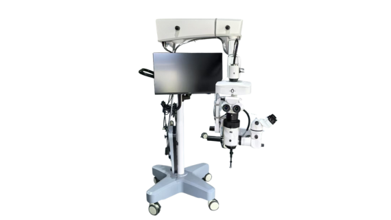
Operating Microscope ROMS100
Characteristics
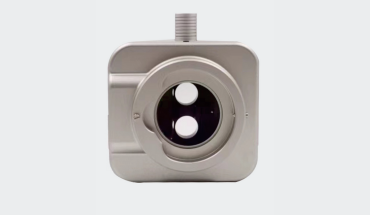
- ROMS 100 Uses Coaxia LED Light Source
- Lighting System Is Equipped with Retina Protection Device, -2° Independent Red Reflex Intensifier, Filter of Infrared
- X/Y System Automatically Controllable, And the Moving Range of X/Y:50mm
- The Instrument Adopts Gas Spring Counter Balance At Every Fixed Point
- The Performance is Easy And Flexible While Stable And Reliable
About Our Ophthalmic Microscope
Our ROMS100 ophthalmic microscope is applicable for both anterior and posterior segment surgeries. The main optical path is designed with four optical paths and adopts a complex chromatic aberration optical system. With Coaxial illumination and two LED lighting sources, our operating microscope provides excellent brightness for surgical procedures.
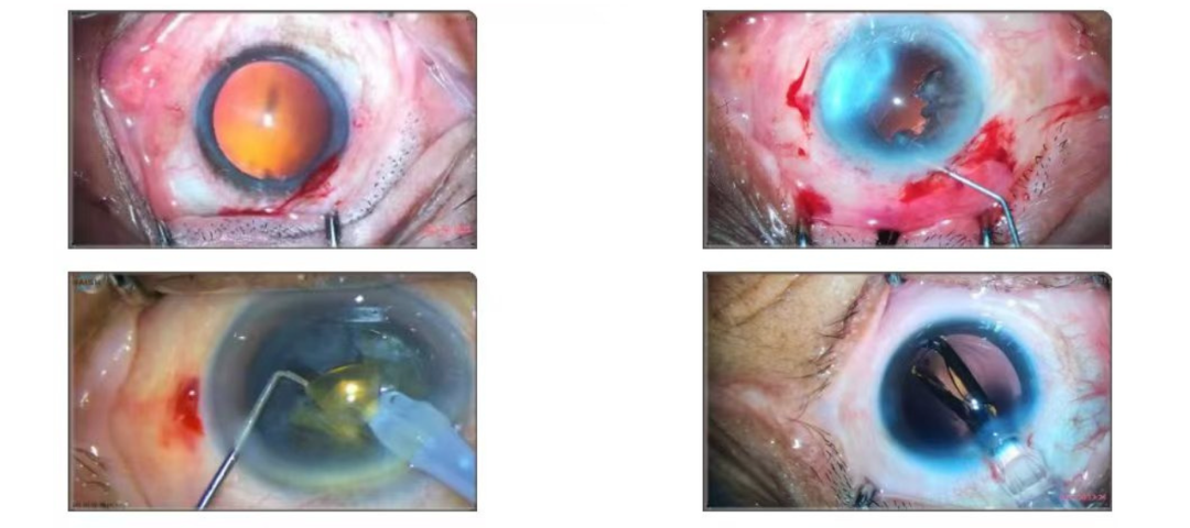
Anterior Segment Surgeries
- You can clearly see the different layers, cloudy areas, and relationships between the surrounding tissues of the crystalline lens.
- Clear surgical area: including surgical incision, anterior chamber, posterior chamber, etc., so that you can accurately perform each surgical step and avoid damaging important surrounding tissues.
- There are no obvious shadow areas within the surgical field of view to ensure that you can observe the surgical site comprehensively.
- The color of the image should reflect the actual situation of the tissue as realistically as possible, so that you can accurately judge the health status and degree of lesions of the tissue.
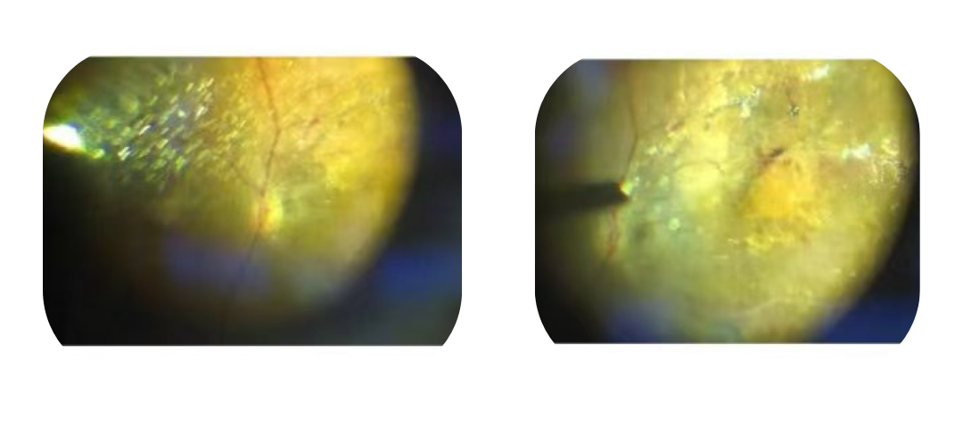
Posterior Segment Surgeries
- Assistant doctors can perform posterior surgeries with high precision under low-brightness lighting conditions.
- Our ophthalmic microscope provides excellent depth of field at 6X to 8X magnification, avoiding frequent focusing adjustments.
- ROMS100 ophthalmic microscope is good at maintaining clear visualization of the retina , especially when using wide-angle 130-degree observation systems.
- During the peeling process, the ROMS100 ophthalmic microscope can also obtain clear visualization of the film layer.
- The design of 4 optical paths ensures sufficient illumination for visualization while avoiding the risk of phototoxic exposure.
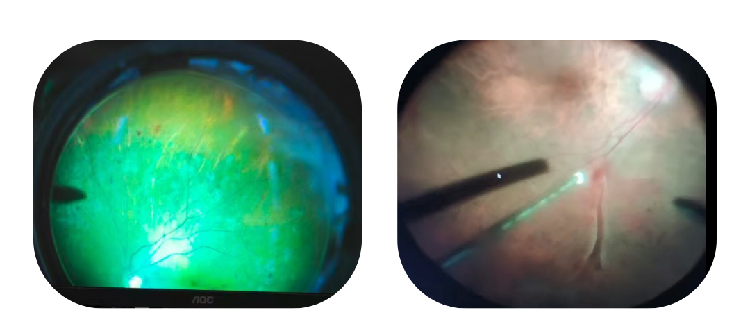
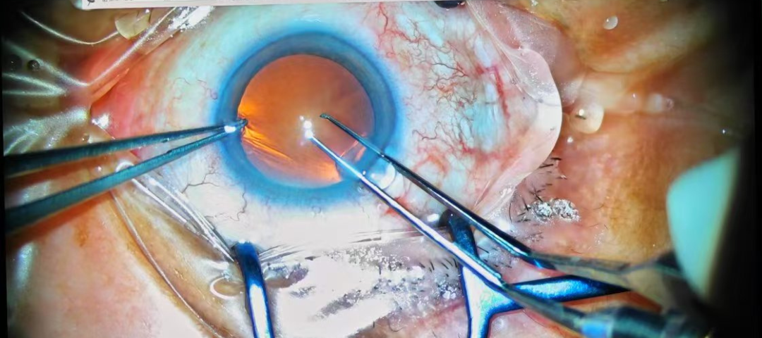
Superior Red Reflex for Retinal Surgeries
The red reflex is a crucial component of ophthalmic surgical microscopes. Its quality directly impacts the microscope’s ability to perform posterior segment surgery. The pupil is illuminated by red light from the microscope, and the reflected red light is observed. Normally, it should appear uniform red. Abnormal color or shape may indicate cataracts, refractive errors, or retinopathy. Our ROMS100 surgical microscope provides clear and bright red reflex, creating an excellent operating environment for posterior segment surgery.
Illumination and Electrical Data
| Main Light | Integrated LED, 3500K, CRI≥95 |
| Red Reflex Illumination | -2°Coaxial, LED, 3500K CRI≥90 |
| Voltage Input | 100~240V, 50/60Hz |
| Fuses | 250V, 10A |
| Electrical Safety Standards | IEC60601-1: 2012, Class I |
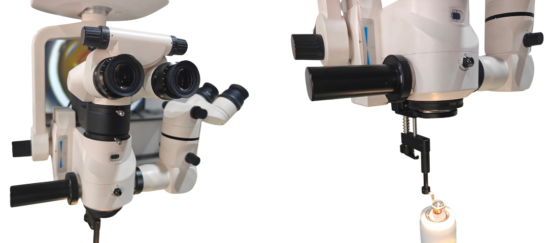
Optical System
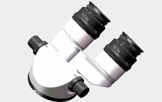
| Eyepieces | 12.5X |
| Diopter Adjustment Range | -7D~+7D |
| Objective | F=200mm |
| Working Distance | 190mm |
| Magnification of Main Optics System | ZOOM 6:1 Motorized |
| Magnifications | Main Optics : 4.1X~24.6X;
Assistant Optics:5.4X, 8X, 12.2X |
| Field Diameter | Main Optics: 8mm~50mm
Assistant Optics: 34.6mm, 23mm, 14.63mm |
| PD For Main Binocular | 51mm~75mm |
| PD For Assistant Binocular | 55mm~75mm |
| Tilt Range of Main Binocular | 0~180° |
| Focusing | 50mm, Motorized with Automatic Reset |
| Focusing Speed | ≤2mm/s |
| Filters | Main Illuminator: Retina Protection Filter, Red-free, GG435 Blue-blocking;
Red Reflex Illuminator:20% Light Attenuation |
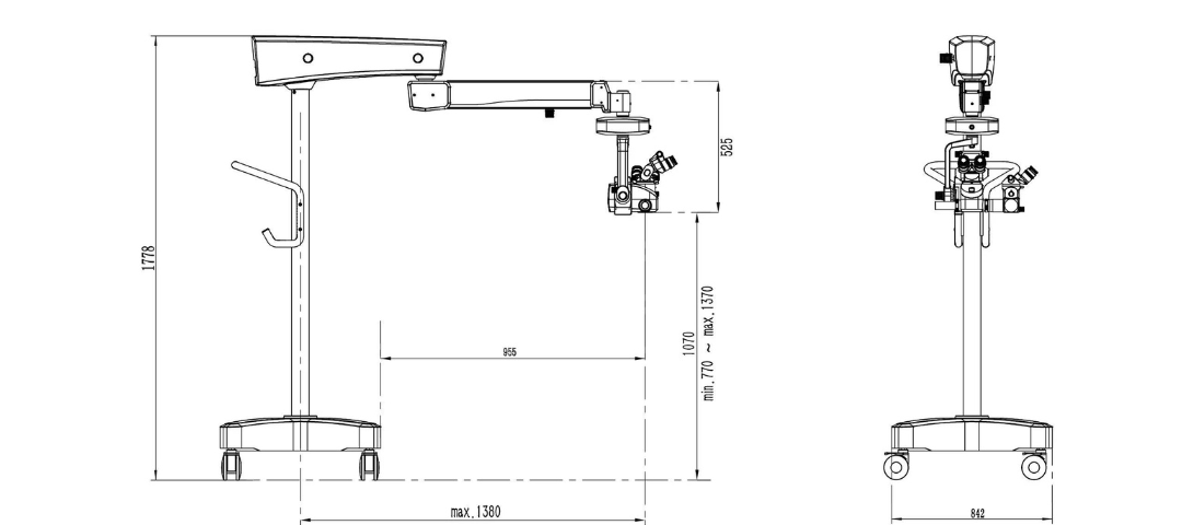
Floor Stand Data
| Foot Wheels | 4*100mm |
| Weight | Approx 185kg, Fully Microscope System |
| Range | Extension 1380mm(Max.) |
| Balancing | Gas Spring |
| XY Unit | 50mm*50mm, Motorized with Automatic Reset |
| XY Moving Speed | ≤2mm/s |
| Tilt Driver | Manual, +15°/-50° |
| Turning Range | Horizontal Arm:360°
Balance Arm:±135° XY Unit:±135° |
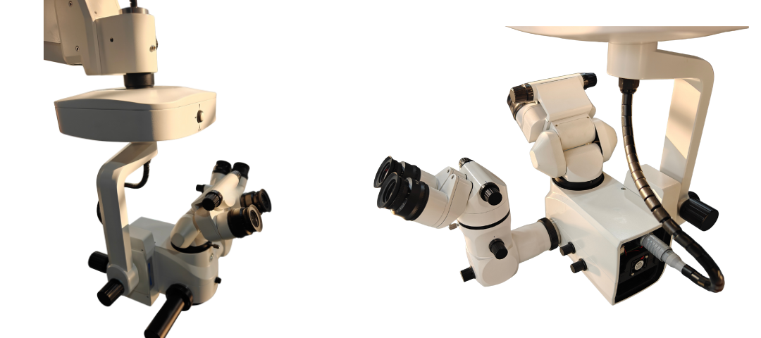
Extendable Scopes for More Operators

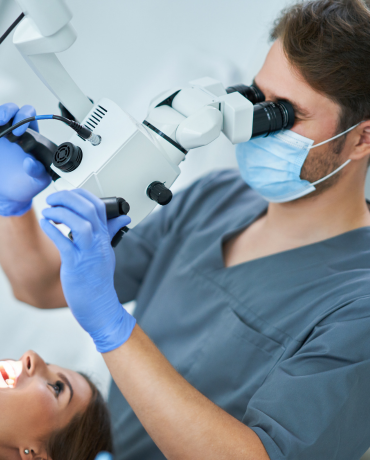
Binocular Tube for 0~180°
The binocular tube of our operating microscope can be adjusted from 0 to 180 degrees, which enhances comfort and flexibility through an even wider range of viewing angles.
Extendable for 2 Operators
An assistant binocular microscope is available with 45-degree inclination, which works in a comfortable standard position with a fixed 45° viewing angle.
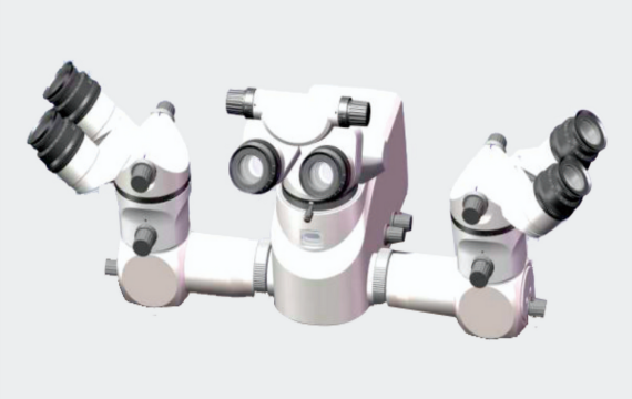
Extendable for 3 Operators
Two assistant scopes are mountable on both sides of the microscope to share the entire surgical process.
Laser Filters
Optional laser protection filters for our ophthalmic microscope provide better protection for the operator, with a protection level greater than 4. There are three wavelength laser filters available: 532nm, 577nm, and 810nm. Welcome to get in touch with us for more information!
Operating Microscope Structure
In the design structure of the surgical microscope, we have fully considered the convenience of the operator and adopted a wired foot pedal to ensure a more stable connection and a smoother surgical process.
Compact and Balanced Microscope
The overall design features the main microscope and an assistant microscope, characterized by a compact and balanced structure that facilitates stable and easy movement.
Reliable Retinal Surgical System
After extensive clinical validation, our posterior segment surgical microscope has gained widespread recognition and high praise. Breaking through technological limitations and achieving a greater depth of field, it provides a superior solution for retinal surgery.
Accessories (3)
-

The integrated 4K HD system is optional, as a digital upgrade system for our ROMS100 surgical microscope, which can be equipped with a high-definition monitor to visualize the surgical process.
-
As an accessory of the surgical microscope, the inverting mirror is used in conjunction with the BIOM system in clinical ophthalmic surgery to invert the inverted retinal image formed by the BIOM to obtain an upright image.
-

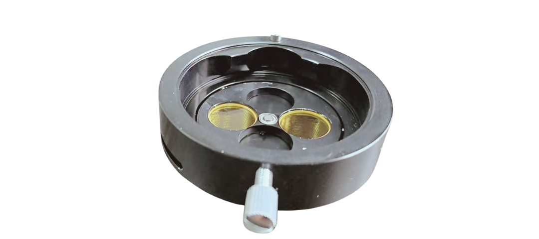
Our BIOM system is an optical system that assists in ophthalmic treatment and surgery through optical principles such as magnification or inversion.
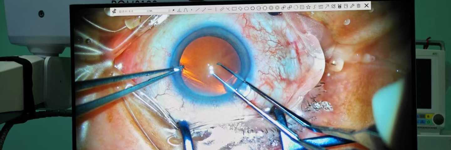
Upgradable 4K Visual System
Our surgical microscope can be upgraded to 4K high-definition system, and when connected to a high-definition monitor, the entire surgical process can be observed in real time. Contact us to upgrade your microscope!
Operating Microscope Application


The ROMS100 microscope is the clearest among domestic brands for posterior segment surgery. When publishing our papers on posterior segment research, we must include the Maxter Medical microscope ROMS100 in the paper.–Prof. Yang from Affiliated Hospital of Qilu University of Technology
Maxter microscope has the clearest optics among the domestic brands I have seen. It is much better than other domestic brands of ophthalmic microscopes I saw at the 2024 National Ophthalmology Annual Meeting. The red light reflex is very good and the depth of field is sufficient for posterior segment surgery, which exceeded my expectations for domestic ophthalmic microscopes.– Prof. He from Hubei Provincial Government Hospital
The ROMS100 microscope is equipped with a 4K high-definition video recording system, which records the entire process of our cataract surgery very well. The images are truly high-definition, which is convenient for us to use for teaching and training.–Prof. Dong from Anhui SuChu Hospital
ROMS100 microscope has the best resolution I never see before for any other Chinese brand ophthalmic microscope, it is really perfect while I check it without lights-turn-on.–Prof. Vadim from TMS Russia