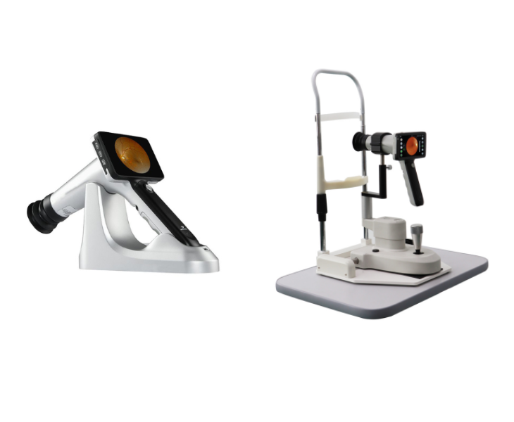
- Es sind sowohl Tisch- als auch tragbare Funduskameras erhältlich
- Nicht mydriatische und sichere Augenuntersuchung
- Wiederaufladbare tragbare Funduskamera
- Mosaik-Fundusbilder
- KI-Diagnose verfügbar
Zuverlässiger Hersteller von Funduskameras
Maxter produziert seit mehr als 30 Jahren Funduskameras 15 Jahre und hat ein ausgezeichnetes R&D-Team, um die kontinuierliche Aktualisierung und Iteration von Funduskameras sicherzustellen. Wir engagieren uns in der Forschung und Entwicklung von Funduskameras ohne Mydriasis. Eine sichere und risikofreie Fundusuntersuchung ist unser ständiges Streben und Ziel.
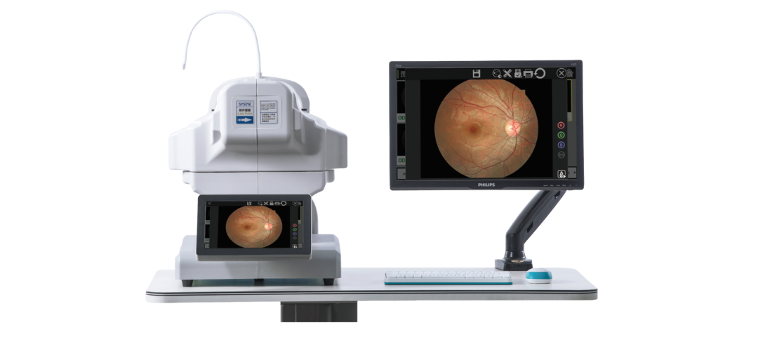
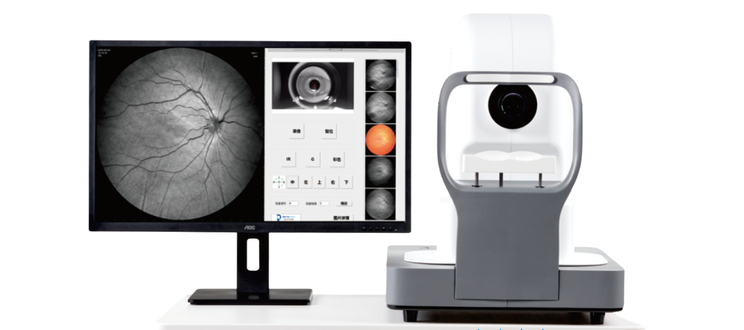
Fundus-Bildgebungssystem
Wir sind auf die Bildgebung des Fundus spezialisiert und legen großen Wert auf eine einfache Bereitstellung, schnell, und effiziente Fundusbildgebung. Von der tragbaren bis zur vollautomatischen Fundusbildgebung, Wir erforschen und forschen ständig, und Aktualisierung unserer Modelle. Bitte bleiben Sie bei unserem Unternehmen auf dem Laufenden, um Updates zu kommenden Funduskameras zu erhalten!


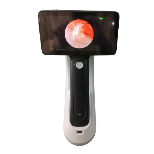
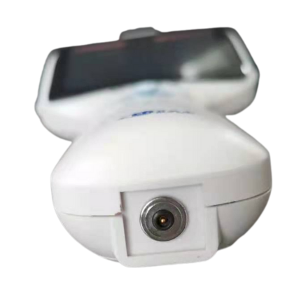
Wertvolle tragbare Funduskamera
Die PFC01 ist unsere kostengünstige tragbare Funduskamera. Es kann sowohl für menschliche als auch tierische Augenuntersuchungen verwendet werden. Es ermöglicht sicherere Fundusuntersuchungen ohne Mydriasis, und kann Pupillen mit einer Größe von nur 2,5 mm erfassen. Die manuelle Fokusfunktion sorgt für Stabilität und Benutzerfreundlichkeit. Diese klassische Handfunduskamera, seit über einem Jahrzehnt ein weltweiter Bestseller, ist Ihre erschwingliche Option.
-
Unsere tragbare Funduskamera hat ein einfaches Design, flexible Tasten, unterstützt die drahtlose Verbindung zu mobilen Geräten, unterstützt Windows- und IOS-Systeme, und ermöglicht eine einfache Datenübertragung.
-
Das innovative Kontaktladedesign erleichtert das Laden, schützt den Ladeanschluss besser, und verlängert die Lebensdauer des Datenkabels
-
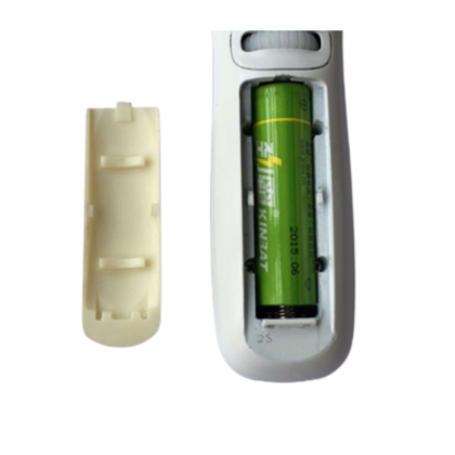
Unsere Funduskamera verfügt über ein wiederaufladbares Design und eine Lithium-Batterie-Stromversorgung, das leicht zu transportieren ist und jederzeit und überall für die Fundusuntersuchung verwendet werden kann. Das Batteriemodell ist 18650, Das ist universell und einfach zu kaufen.
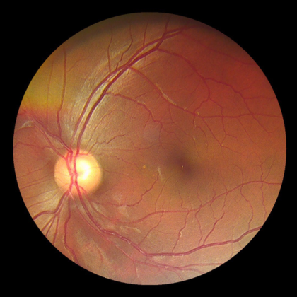
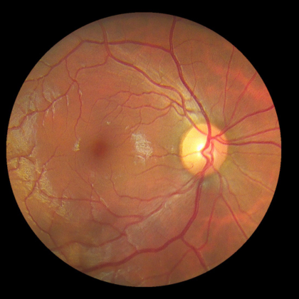
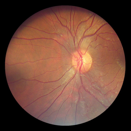
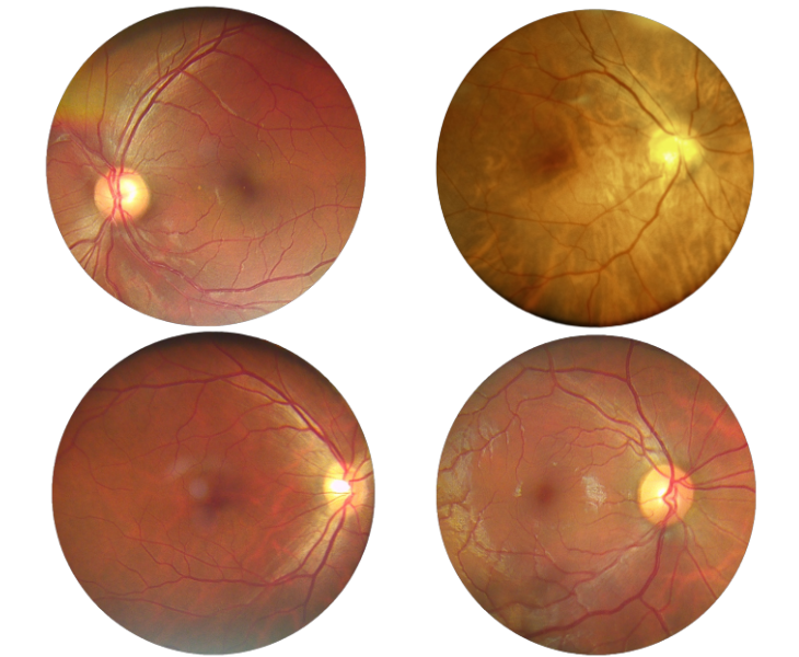
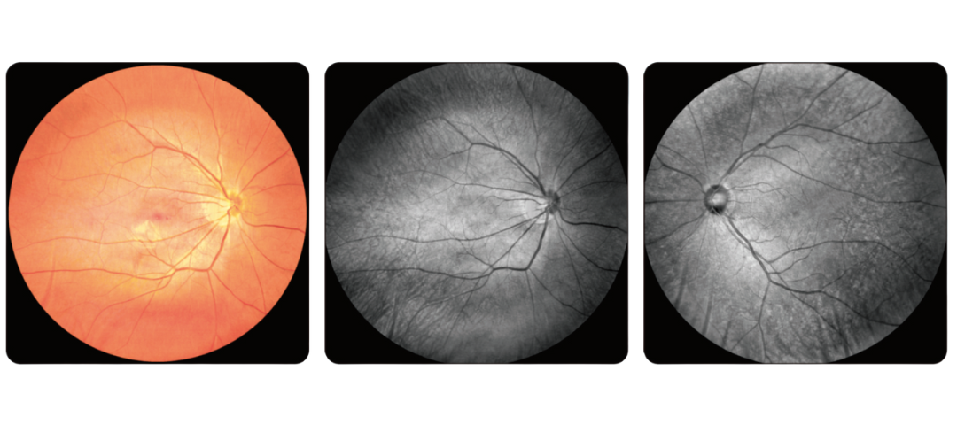
Fundusbilder
Unsere Funduskamera verfügt über ein 45-Grad-Sichtfeld, Ermöglicht die einfache Erfassung hochauflösender Fundusbilder. Im Lieferumfang ist außerdem eine kostenlose Apple-Software enthalten, die eine drahtlose Echtzeitübertragung auf mobile Geräte ermöglicht.
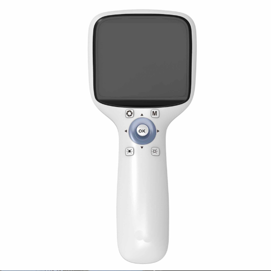
Tragbare Premium-Funduskamera
Die PFC11 ist unsere tragbare Premium-Funduskamera. Es verfügt über fünf interne Fixierungslichter, 18 Megapixel, ein Android-Betriebssystem, und ein integriertes System zur Verwaltung medizinischer Unterlagen. Es wird mit einer leichten und robusten Tragetasche geliefert, So können Sie jederzeit Fundusuntersuchungen durchführen, überall. Jetzt, Kontaktieren Sie uns, um Ihre eigene mobile Funduskamera zu kaufen!
50° Sichtfeld
Die hochwertige tragbare Funduskamera bietet ein 50-Grad-Sichtfeld, So können Sie den Fundus in einem größeren Bereich sehen. Es unterstützt die intelligente KI-Diagnose, Autofokus, und automatische Fotoaufnahme. Die optionale Halterung verwandelt sie im Handumdrehen in eine Desktop-Funduskamera.
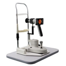
5 Megapixel-Funduskamera
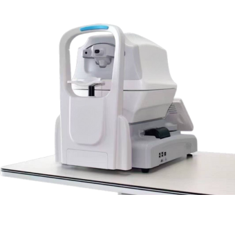
Wir haben diese 3100 m große, vollautomatische, nicht-mydriatische Funduskamera auf den Markt gebracht, das ein Dual-Kamera-Erfassungssystem verwendet und die kleinste nicht-mydriatische Pupille von bis zu 2,8 mm erfassen kann. Diese Funduskamera verfügt über eine Soft-Exposure-Technologie, Dadurch wird die Belichtungshelligkeit reduziert, um den Patientenkomfort während der Untersuchung zu erhöhen.
![]()
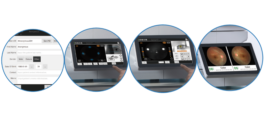
Automatischer Arbeitsmodus
Die 3100-m-nicht-mydriatische Funduskamera verfügt über automatische und manuelle Arbeitsmodi. Im Automatikmodus, Sie müssen lediglich ein Patientenprofil erstellen, und Sie können die Kinnstütze automatisch anpassen, automatisch fokussieren, automatisch ausrichten, und mit einem Klick automatisch Bilder aufnehmen.
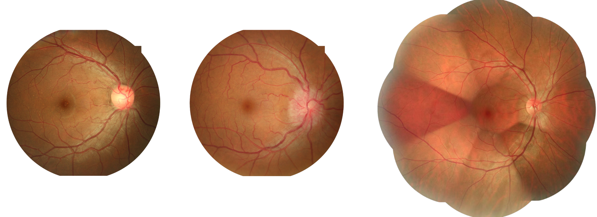
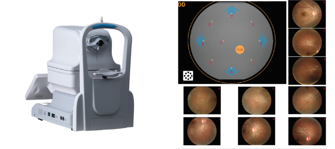
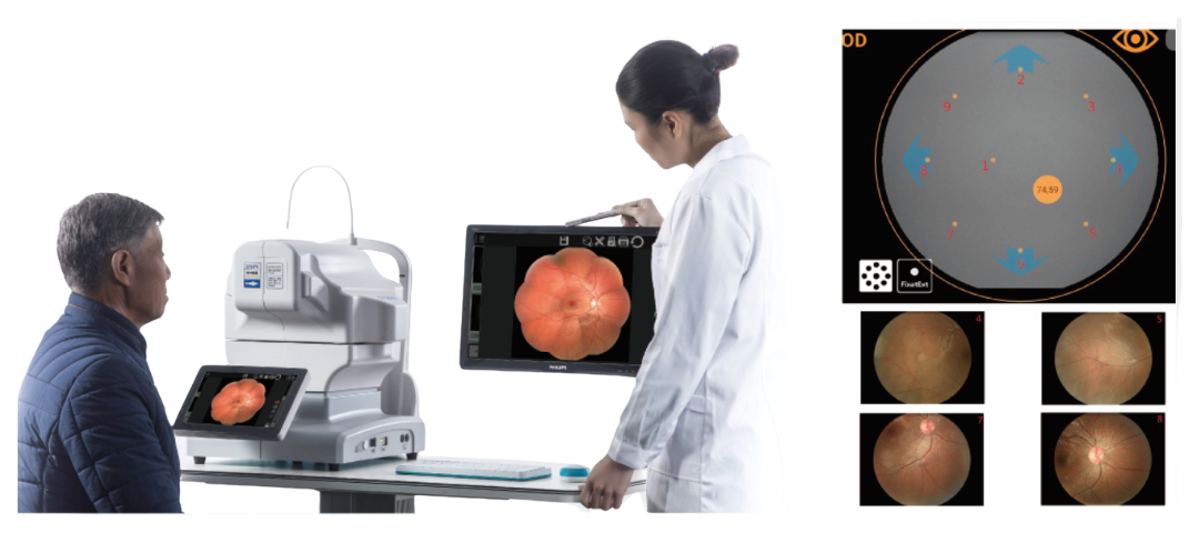
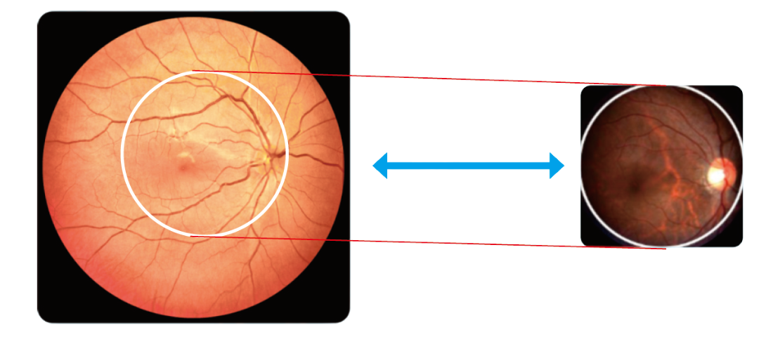
135 Gradweites Feld
9 Fundusbilder unter 50 Grad kann zu einem 135-Grad-Ultraweitwinkel kombiniert werden, um den gesamten Fundus zu sehen. Die Funduskamera hat 9 Zielfixierungen, und mehr Fixierungen werden zwischen den verteilt 9 interne Fixierungen dargestellt.
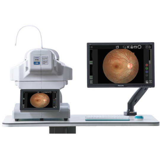
24 Megapixel-Funduskamera
Das ist unser Vielseitigkeitsmodell, nicht mydriatisch, vollautomatische Funduskamera. Seine 24-Megapixel-Kamera erfasst jedes Detail des Fundus in hoher Auflösung. Es verfügt über ein 50-Grad-Sichtfeld und unterstützt sowohl den mydriatischen als auch den nicht-mydriatischen Modus.
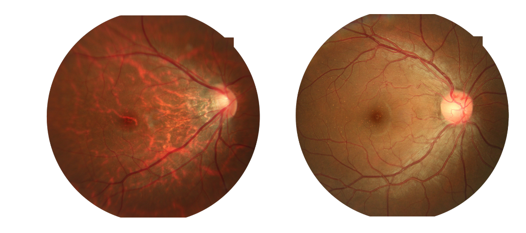
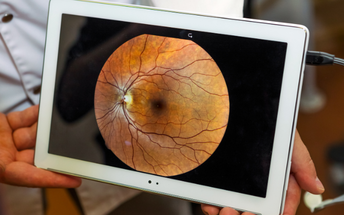
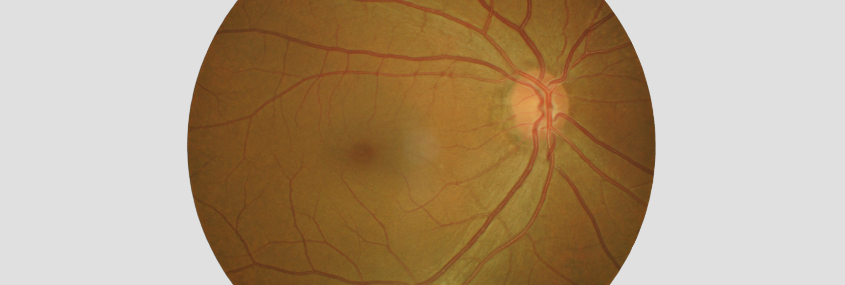
Hochauflösendes Fundusbild in jedem Detail
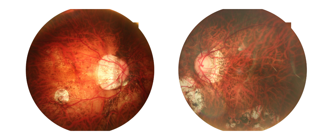
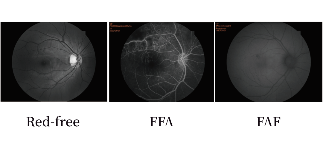
Funduskamera mit FFA
Das 3100 Die vollautomatische Funduskamera verfügt optional über Rotfrei, FFA, FAF, und andere Funktionen. Es kann farbige Fundusbilder erfassen und auch die Anforderungen der Fluoreszenzangiographie erfüllen. Es handelt sich um eine kostengünstige Funduskamera.
Ein Klick zur großen Feldansicht bis zu 135°
Mit einem intelligenten Krankenaktenverwaltungssystem und One-Touch-Bedienmodus, Unsere vollautomatische Funduskamera bietet weitere Funktionen zur Fundusuntersuchung, Optimieren Sie Ihren Arbeitsablauf und verbessern Sie die Effizienz. Entdecken Sie mit uns weitere Wunder des Fundus!
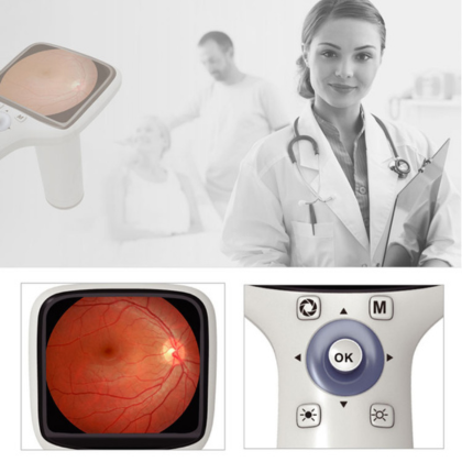
Nicht-mydriatische Funduskameraanwendungen
Unsere modernen Funduskameras digitalisieren Fundusdaten. Dies ermöglicht eine Archivierung, Lagerung, Teilen, und Fernberatung. Außerdem, Sie können in KI-gestützte intelligente Diagnosesysteme integriert werden, um zuverlässige Diagnoseergebnisse zu liefern.
Mit der Weiterentwicklung moderner Augenkrankheiten und der zunehmenden Beliebtheit der Laser-Augenchirurgie, Die postoperative Nachsorge ist ein wesentlicher Bestandteil der postoperativen Genesung. Wir können maßgeschneiderte Lösungen für die postoperative Nachsorge anbieten.
Technischer Parametervergleich
Vergleich der Parameter tragbarer Funduskameras
| Tragbare Funduskamera | PFC01 | PFC11 |
| Fov | 45 Grad | 50 Grad |
| Blitzmodus | Weiße LED/ Nahinfrarot -LED | Weiße LED/ Nahinfrarot -LED |
| Auflösung | 1920*1080 | 12 Megapixel |
| Min. Schüler | 2.5mm | 2.5mm |
| Fokus | Handbuch | Maunal / Rehtta |
| Fixierung | NEIN | 5 Interne Fixierungen |
| Bildschirm | 3.5-Zoll-TFT-Farbbildschirm | 4.7-Zoll Multi-Touch-Bildschirm |
| Format | JPEG | Farbbild, Monochromes Bild, Infrarotbild |
| Schnittstelle | Micro-USB | USB3.0 |
| Bildübertragung | Micro-USB, W-LAN | USB3.0, WiFi, Bluetooth,SD, Typ c |
| Lagerung | Micro-SD-Karte, maximal 32G | 4G+64G |
| Leistung | Li-on-Akku 3,6 V/3400 mAh | Li-on-Akku 3,7 V/3500 mAh |
| Nettogewicht | 400G | 600G |
| Größe | 19*10*21cm | 41*25*35mm |
Vergleich der Parameter der Auto-Funduskamera
| Automatische Funduskamera | 3100 | 3100M |
| Erfassungsmodi | Nicht-Mydriatisch/Mydriatisch
Vordere Fotografie Rotfrei(Optioanl) FFA(Optional) FAF(Optioanl) |
Nicht-Mydriatisch/Mydriatisch |
| Sichtfeld | 50° | 50° |
| Arbeitsabstand | 35mm | 13mm |
| Mindestschülergröße | ≥mm3,3 | ≥2,8 mm |
| Fokusmodus | Automatisch/manuell | Automatisch/manuell |
| Belichtungsmodus | Automatisch/manuell | Automatisch/manuell |
| Betrieb | Automatisch/manuell | Automatisch/manuell |
| Fotografie | Spiegelreflexkamera | Spiegelreflexkamera |
| Auflösung | 24Megapixel | 5 Megapixel |
| Dioptrienausgleich | ±25D | ±25D |
| Fixierung | Interne Fixierung/externe Fixierung | Interne Fixierung |
| Autofokus-Unterstützung | Infrarot geführt | Dual-Kamera |
| DICOM 3.0 | Ja | Ja |
| Anpassung AI-Port | Ja | Ja |
| Abmessungen | 38*56*48cm | 28*56*46cm |
| Gewicht | 27kg | 21kg |
| Stromversorgung | 100-240V 50/60Hz | 100-240V 50/60Hz |
-
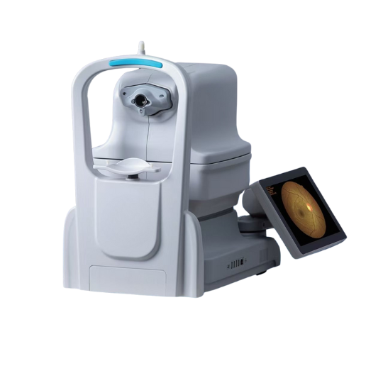
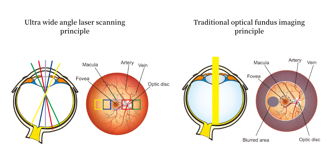
Konfokales Laserscannen
Die konfokale Ultraweitfeld-Laser-Funduskamera der neuen Generation unterstützt nicht-mydriatische und Ultraweitfeld-Bildgebung. Der Einsatz eines konfokalen Lasersystems ermöglicht schnelle und präzise Untersuchungen, Verbesserung des Patientenkomforts, insbesondere für Patienten mit Glaukom oder Diabetes, die für eine Mydriasis ungeeignet sind.
Weitfeld-Netzhautbildgebung
Unsere konfokalen Laser-Funduskameras unterstützen mehrere Lichtquellenoptionen, Dies ermöglicht die einfache Erzielung eines Ultraweitwinkel-Sichtfelds von 130° und 168°, und Ermöglichen der Bilderfassung mit einem Klick. Momentan, Für die Aderhautbeobachtung ist die Betrachtung mit einer Lichtquelle möglich, während die Betrachtung mit zwei Lichtquellen sowohl für die Netzhaut als auch für die Aderhaut möglich ist. Ein Quad-Lichtquellen-Betrachtungssystem für die Netzhaut und die Aderhaut ist bald verfügbar.
SLO-Arbeitsprinzip
Netzhaut-Vollschichtfotografie + Video
Die vollschichtige Bildgebungs-/Videografietechnologie der Netzhaut ermöglicht eine genauere Identifizierung pathologischer Bereiche, Bereitstellung einer Grundlage für eine individuelle Behandlung.
Normalerweise, Wir haben Produkte auf Lager, und wir können nach innen versenden 2 Werktage nach Erhalt Ihrer Zahlung! Bitte kontaktieren Sie uns für die genaue Lieferzeit für Großbestellungen!
Ja, Bei Bedarf können wir Schulungen anbieten! Bitte kontaktieren Sie uns für unser Schulungsvideo!
Allgemein, Wir gewähren 1 Jahr Garantie auf unsere Produkte. Aber für einige Modelle, Wir bieten eine längere Garantiezeit. Kontaktieren Sie uns für weitere Informationen zur Garantiezeit!