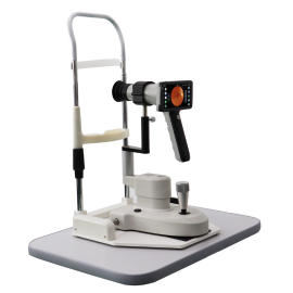
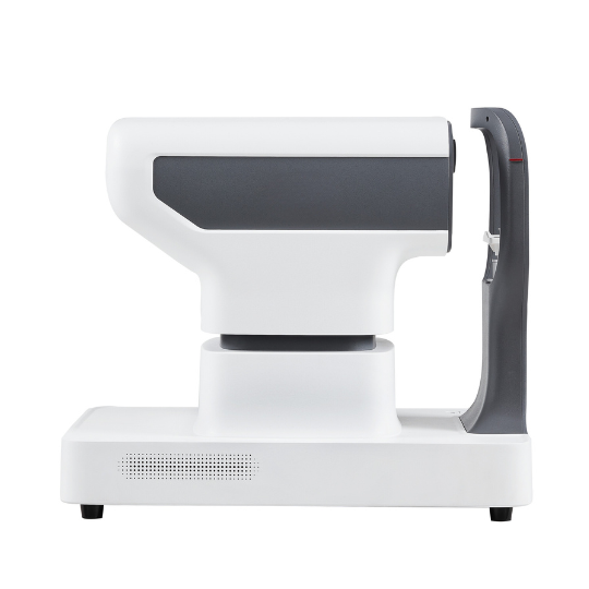
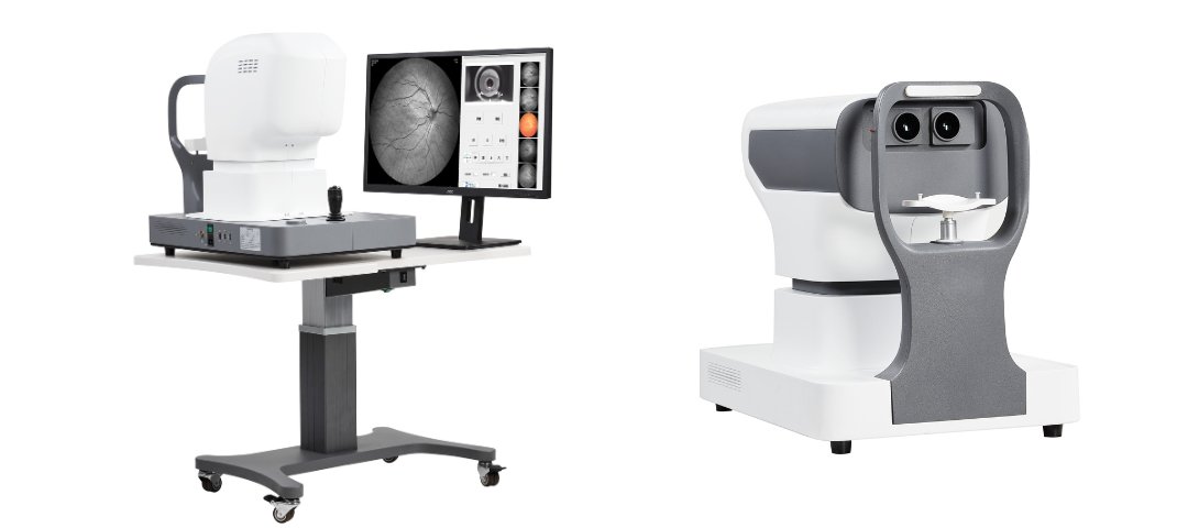
- Superior imaging quality and field of view
- Lowest price laser fundus camera
- Supports AI diagnosis
- Multi-laser option
- 130 degrees and 168 degrees are available
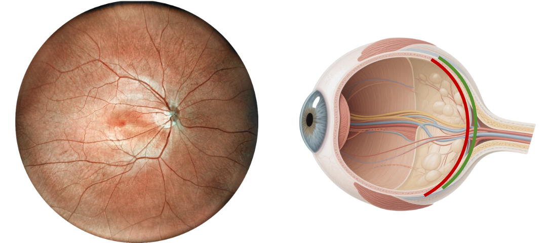
Advantages of Laser Scanning Light Source
The light source of a fundus camera determines its effectiveness. The LED light used in optical fundus cameras is highly irritating, has a narrow range, and lacks penetration. The laser light source used in the laser fundus camera has low stimulation, a wider field of view, and strong penetration, enabling in-depth observation. Laser fundus imaging provides richer information and a more accurate diagnosis.
Parameters Comparison
| Optical Fundus Camera | Laser Fundus Camera | |
| Irradiation stimulation | 3J/s | 3 mJ/s |
| Imaging range | Upper limit 80 degrees | Starting from 100 degrees |
| Deep imaging due to penetration | Retina | Retina and choroid |
| Operation Mode | Manual+Auto | Electric control + Auto focus and shooting |
| Working Environment | Dark room | Anywhere |
| Video | No | Yes |
| Mydriasis | Most need | No |
| Composite Image | Some | Support 2-5 stitched images, with a maximum range of 220 degrees or 270 degrees |
Optical Fundus Camera Get Retinal Layer
Traditional optical fundus cameras have a small field of view, generally 40 to 60 degrees, and can only see the retinal layer of the fundus, but cannot obtain a complete fundus image.
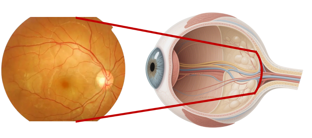
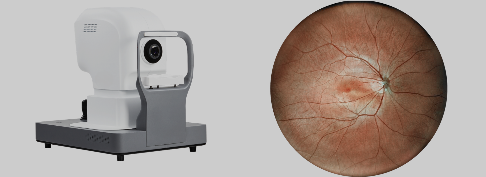
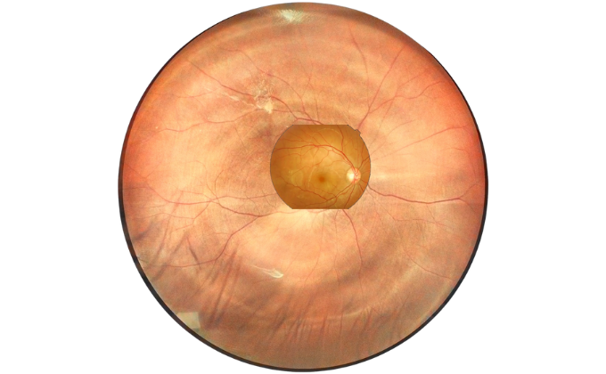
Scanning Laser Ophthalmoscope Obtain Retinal Layer and Choroidal Layer
Our scanning laser ophthalmoscope uses multi-wavelength laser scanning to penetrate the fundus, capture details of the retinal and choroidal layers, and obtain high-definition fundus images.
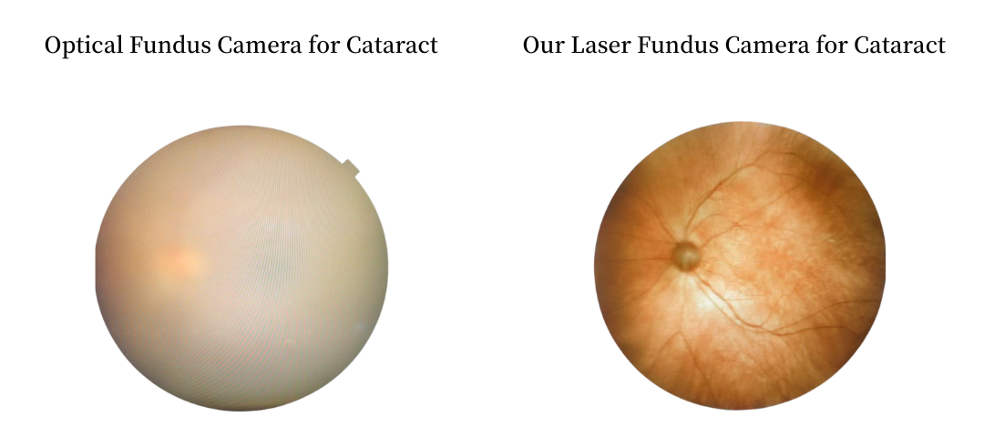
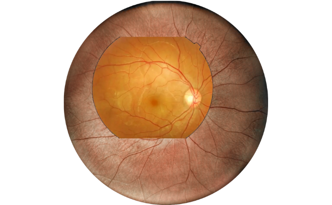
Retina Examination Under Cataracts
Examining fundus diseases through cataracts is a crucial topic in ophthalmology. Traditional fundus cameras cannot see through cataracts to capture fundus conditions. However, our DF600 scanning laser ophthalmoscope can penetrate moderate cataracts, providing a glimpse into the fundus and offering greater convenience for fundus examinations in patients with cataracts.
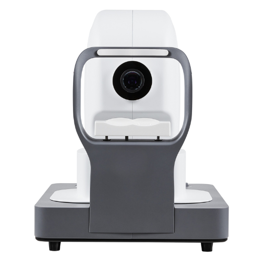
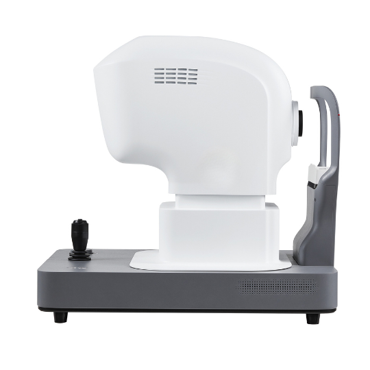
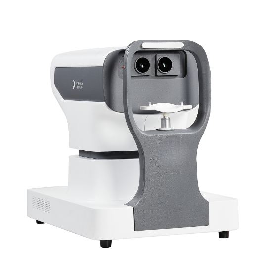
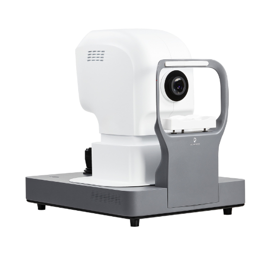
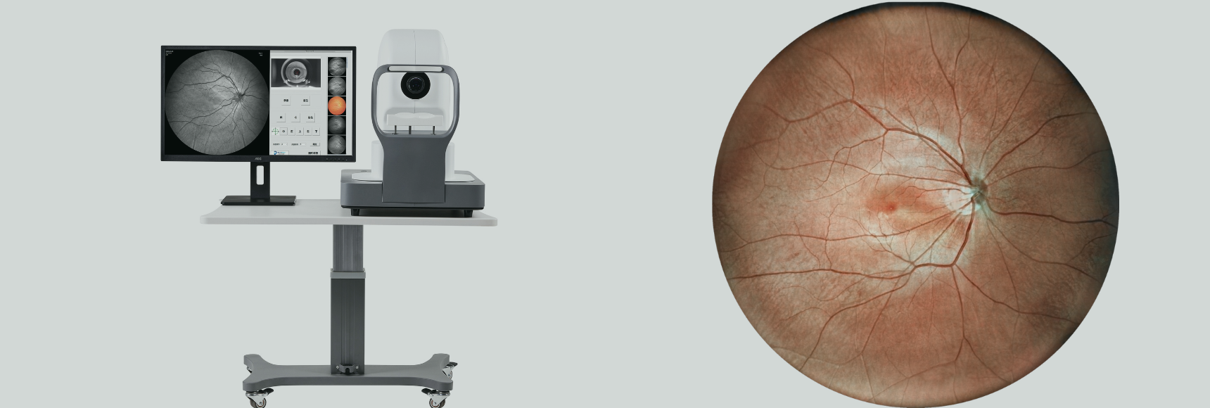
DF600 Configuration
The DF600 fundus camera is available in multiple configurations. Two wide-angle options are available: a 130-degree and a 168-degree field of view. Different numbers of laser light sources can be used to observe various parts of the fundus. Please choose the appropriate light source according to your needs.
| Field of View | 130 Degree | 168 Degree |
| Single Laser | Retina | Retina |
| Double Lasers | Retina and choroid | Retina and choroid |
| Four Lasers | Retina and choroid | Retina and choroid |
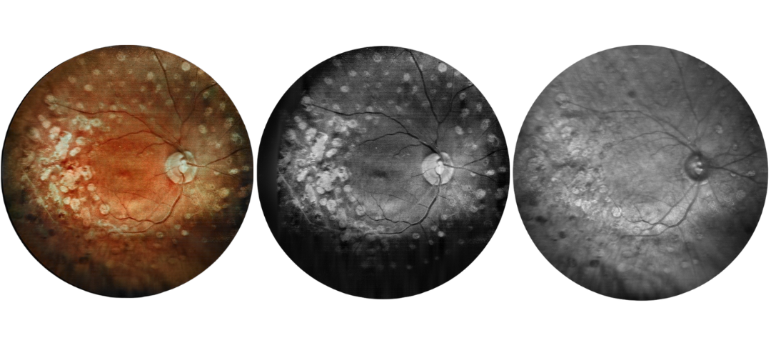
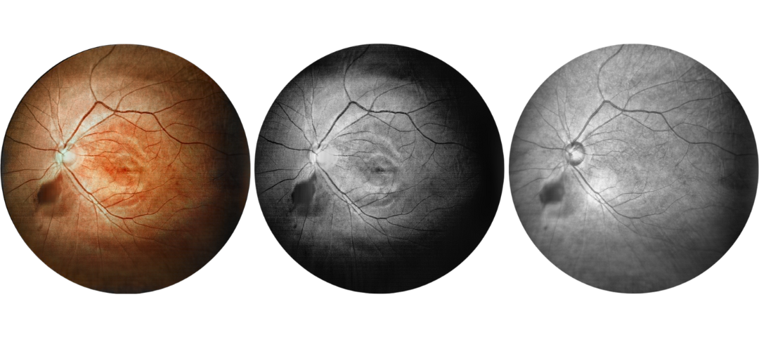
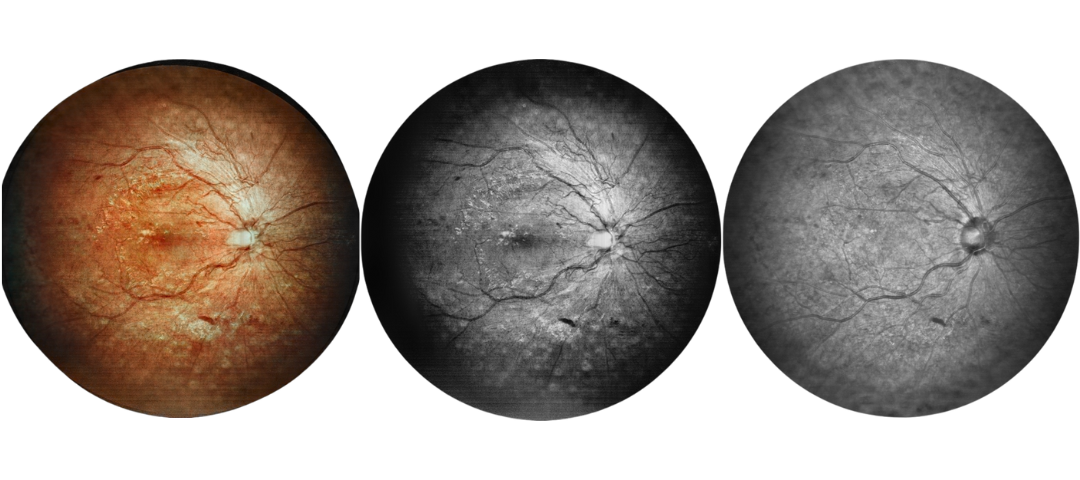
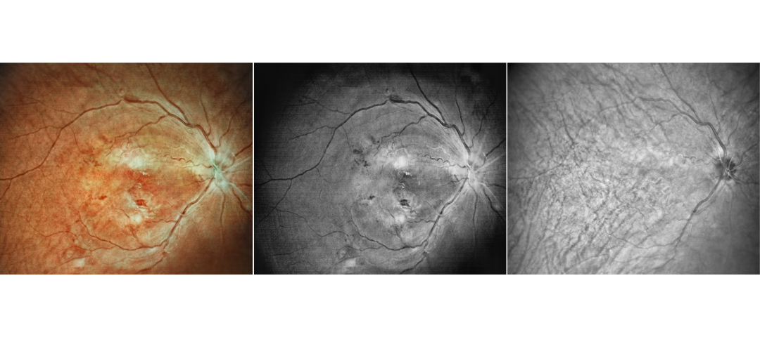
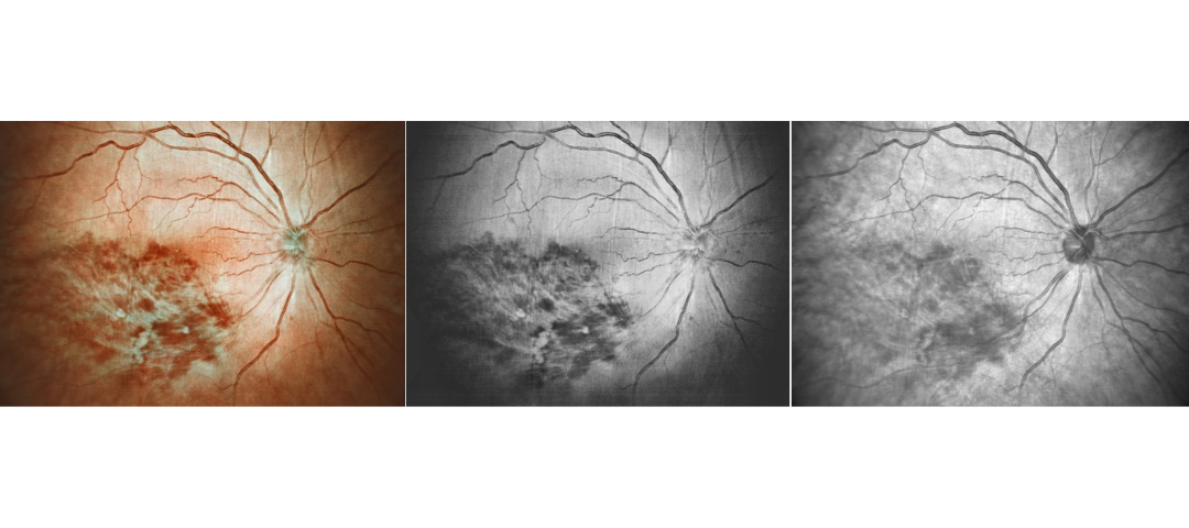
Fundus Cases Display
Our SLO makes fundus examination easier for you, capturing ultra-clear and wide-angle images of the retina and choroid, and then synthesizing color fundus images to present a rich picture of the fundus status.
Diabetic retinopathy: post photocoagulation
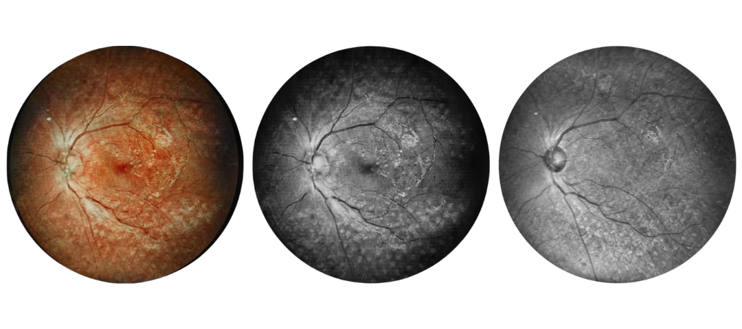
Diabetic retinopathy: bleeding and microvascular lesions
Diabetic retinopathy: post photocoagulation
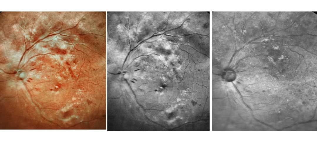
Diabetic retinopathy: bleeding, exudation, and post-photocoagulation
Diabetic retinopathy: bleeding
Diabetic retinopathy: bleeding and exudation
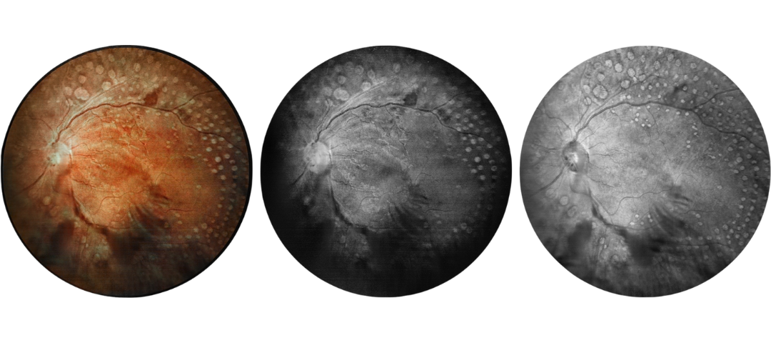
Diabetic retinopathy: vascular abnormalities, bleeding spots, and post-photocoagulation
Diabetic retinopathy: bleeding, exudation and choroidal vascular abnormalities
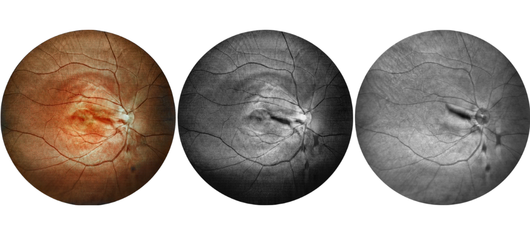
High blood pressure, venous obstruction, and bleeding
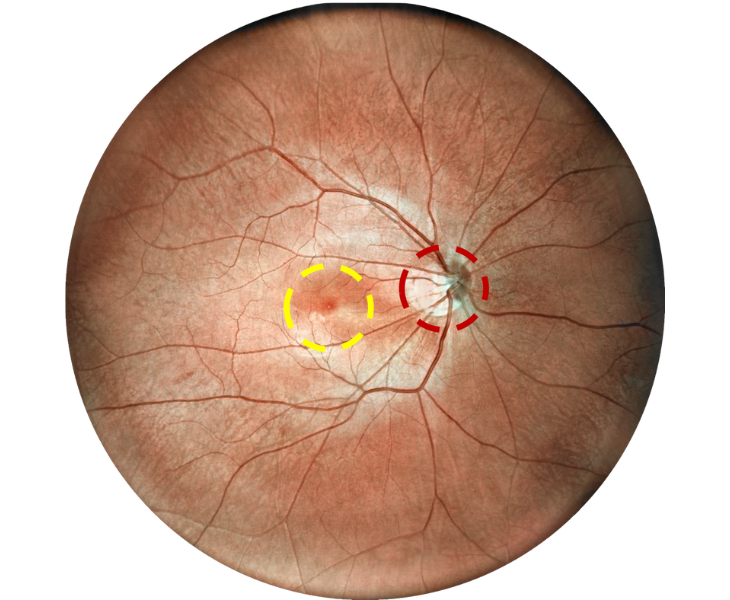
Observation Range of SLO
| Observation Area | Single Laser | Double Laser | Four Laser |
| Optic Disc | Clear | Clear | Clear |
| Vessels | Clear | Clear | Clear |
| Macula | Unobservable | Clear | Clear |
| Fundus tissue | Clear | Clear | Clear |
Ultra Wide Field of View
An 80-degree optical fundus image and a 130-degree laser fundus image, a more intuitive comparison, help you choose the fundus camera that suits you best.
Compared to 60-degree optical fundus images, our 168-degree laser fundus camera shows more dimensions and details, helping you make a more accurate diagnosis.
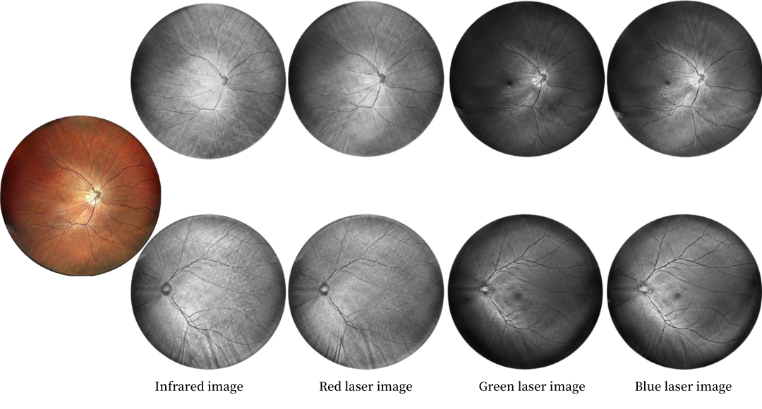
Multi-wavelengh Laser Imaging
Single Infrared Laser
Infrared laser wavelength 840 for retina imaging at 130 degrees
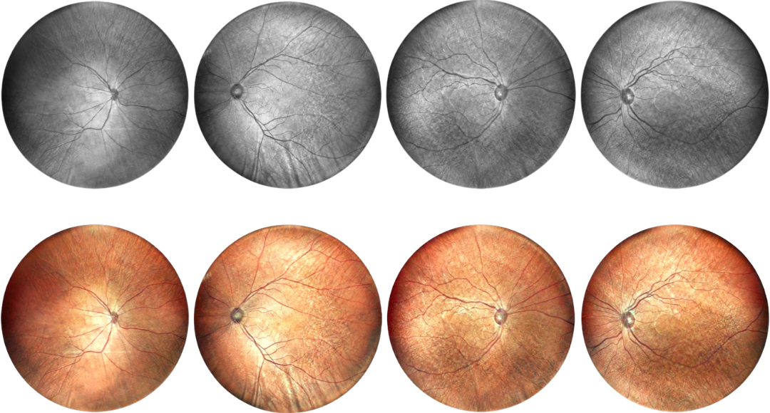
Double Lasers
Infrared laser 840 wavelength and 520 green laser for retina and choroid imaging at 130 degrees
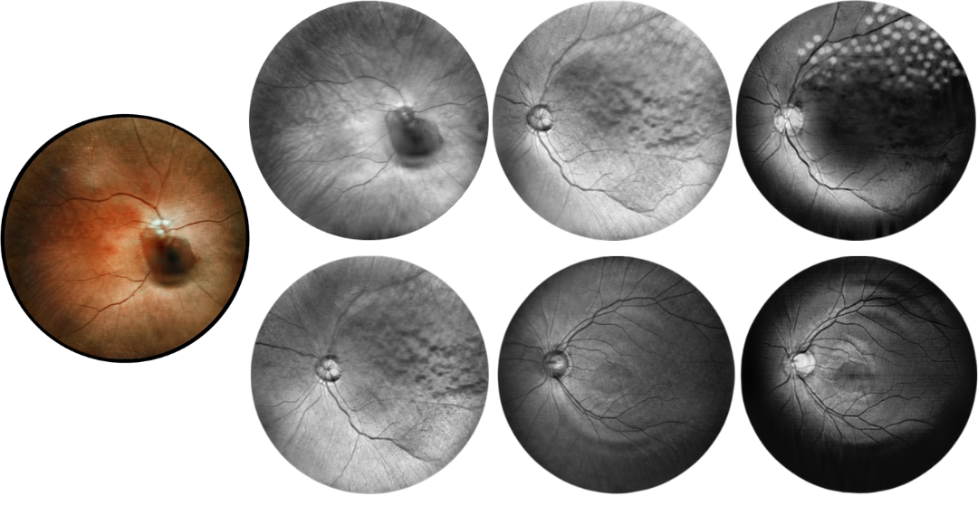
Four Lasers
Infrared laser and green+red+blue laser for retina and choroid imaging at 130 degrees
-
AI Detection System
Our company’s DF600 SLO camera, equipped with an AI-assisted fundus image detection system, can efficiently identify the following five types of fundus diseases. This system helps users quickly screen for fundus diseases, clarify the scope of further examination, and reduce missed diagnoses. It also enhances work efficiency, ensures a quality review, and reduces the difficulty of image interpretation, making it easier for non-specialists to perform preliminary image analysis.
Customizable Report
Reports can be customized and uploaded to the hospital’s internal system, making image interpretation intuitive and processing extremely fast. It is suitable for large-scale examinations in various scenarios.
The AI-assisted detection system classifies fundus diseases into the following five categories:
- Abnormal black spots
- Abnormal macula
- Abnormal blood vessels
- Abnormal optic disc
- Abnormal fundus structures
Normally, we have products in stock, and we can ship out within 2 working days after receiving your payment! Please contact us for the exact lead time for bulk orders!
Yes, we can offer training if you need! Please get in touch with us for our training video!
Generally, we offer a 1-year warranty for our products. But for some models, we offer a longer warranty period. Contact us for more information about the warranty time!